. 2016 Feb;11(2):193-213.
doi: 10.1038/nprot.2016.009. Epub 2016 Jan 7.

In vitro reconstitution of B cell receptor-antigen interactions to evaluate potential vaccine candidates

Affiliations

In vitro reconstitution of B cell receptor-antigen interactions to evaluate potential vaccine candidates

Grant C Weaver et al. Nat Protoc. 2016 Feb.

Abstract

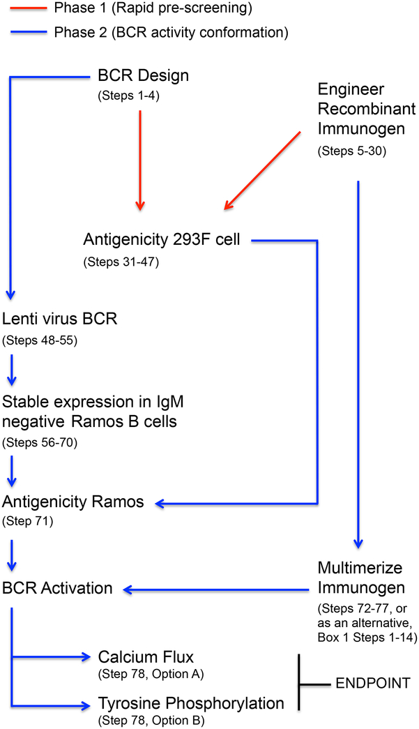
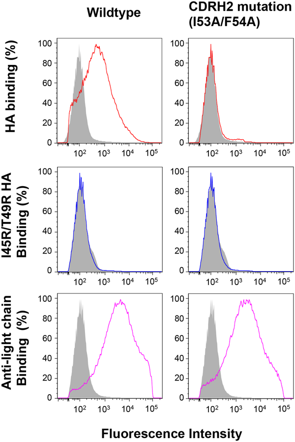
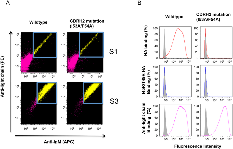
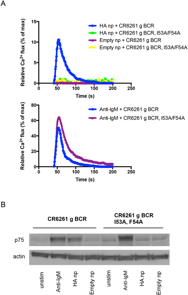
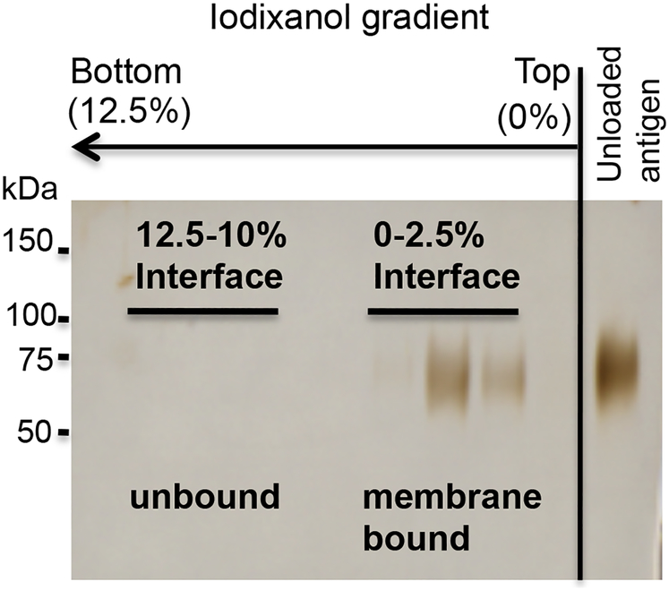
Predicting immune responses before vaccination is challenging because of the complexity of the governing parameters. Nevertheless, recent work has shown that B cell receptor (BCR)-antigen engagement in vitro can prove a powerful means of informing the design of antibody-based vaccines. We have developed this principle into a two-phased immunogen evaluation pipeline to rank-order vaccine candidates. In phase 1, recombinant antigens are screened for reactivity to the germline precursors that produce the antibody responses of interest. To both mimic the architecture of initial antigen engagement and facilitate rapid immunogen screening, these antibodies are expressed as membrane-anchored IgM (mIgM) in 293F indicator cells. In phase 2, the binding hits are multimerized by nanoparticle or proteoliposome display, and they are evaluated for BCR triggering in an engineered B cell line displaying the IgM sequences of interest. Key developments that complement existing methodology in this area include the following: (i) introduction of a high-throughput screening step before evaluation of more time-intensive BCR-triggering analyses; (ii) generalizable multivalent antigen-display platforms needed for BCR activation; and (iii) engineered use of a human B cell line that does not display endogenous antibody, but only ectopically expressed BCR sequences of interest. Through this pipeline, the capacity to initiate favorable antibody responses is evaluated. The entire protocol can be completed within 2.5 months.


Conflict of interest statement

Competing financial interests The authors declare that they have no competing financial interests.

Figures

Figure 1.
Figure 1.
BCR antigen recognition pipeline. This protocol is used to assess whether candidate immunogens can engage and stimulate BCRs of interest, namely germline antibody sequences that give rise to effective humoral responses following infection with pathogen. The protocol is divided into two phases: 1) a rapid screening step wherein antigen interactions with low-affinity germline mIgM are evaluated at the 293F cell surface; and 2) a subsequent analysis of the capacity to stimulate BCR activity through the engagement specificity identified in Phase 1.
Figure 2.
Figure 2.
Inferring a germline reverted antibody. Presented is an example using the heavy chain variable region from CR6261, an IGHV1–69-derived bnAb against influenza virus,. The nucleotide sequence for the variable region (FR1, CDR1, FR2, CDR2, FR3, CDR3, FR4) was entered into JoinSolver and analyzed using the Kabat nomenclature setting. The two important features of the output are: 1) the highest scoring V gene alignment (=V gene origin); and 2) the position and sequence of the mature CDR3. Information on the D and J genes used to generate the CDR3 are also provided in the output, but they are not displayed or used in this protocol as this junctional information it is often not clear and is consequently not standard to germline reversion.
Figure 3.
Figure 3.
A leader sequence should precede the variable region of germline-reverted antibody. This will insure that the mIgM ectodomain is assembled in the lumen of the ER. For CR6261, the leader sequence “mdwtwrflfvvaaatggvqs” (underlined) is used. Inputting the leader + variable region sequence into the SignalP 4.1 server (http://www.cbs.dtu.dk/services/SignalP/) confirms the presence and position of the introduced signal peptide cleavage site.
Figure 4.
Figure 4.
Overlap PCR to generate membrane anchored IgM configuration of germline reverted antibodies. Two fragments, the variable region (+leader sequence, underlined) and mIgM constant region are first amplified by individual PCR. Primers for these reactions are displayed in color. We recommend a minimum of 20bp overlap for the design of primers 3 and 4. Following amplification and gel purification of the two fragments, they are combined and subject to another round of PCR using only primers 1 and 4. The resultant combined fragment should run at ~1.8 kb. Primers 1 and 4 should be designed with the appropriate restriction enzyme sites to ligate the combined product into the desired expression vector.
Figure 5.
Figure 5.
Membrane presented antibody as a means to pre-screen candidate immunogens for the capacity to engage germline BCR sequences of interest. Candidate immunogens are fluorescently labeled and their binding to mIgM antibody expressed on the surface of 239F cells is evaluated by flow cytometry. Presented is CDRH2-dependent recognition of the influenza HA stem region by germline CR6261 (colored lines; grey is binding to 239F expressing VRC01 mIgM, an irrelevant antibody sequence). Surface trafficking of both wildtype and CR6261 germline mIgM containing a I53A/F54A mutation in CDRH2 is confirmed using a fluorescent anti-light chain antibody. Specificity for the HA stem is defined by reactivity to wildtype HA but not HA containing the mutation I45R/T49R in the stem epitope.
Figure 6.
Figure 6.
Lentiviral mediated expression of BCR in Ramos. To assess BCR activity in response to antigen, heavy and light chain components are expressed in a clone of Ramos expressing no endogenous mIgM at the cell surface. Following enrichment by FACs (A), BCR antigenicity (B) is assessed by flow cytometry. CDRH2-dependent recognition of the influenza HA stem by germline CR6261 (colored lines) versus irrelevant IgM VRC01 IgM BCR (grey) is assessed by binding of fluorescent HA (but not I45R/T49R HA) to wildtype BCR but not to its mutant form containing a I53A/F54A substitution in CDRH2. S1 refers to the first sort after lentiviral infection and S3 shows the cells after three rounds of BCR sorting/enrichment.
Figure 7.
Figure 7.
BCR activation in response to multimerized candidate immunogen. For a kinetic readout, multimerized antigen is added to cells displaying BCR of interest that have been equilibrated with Fura Red, a ratiometric calcium-reporting fluorescent dye. Calcium bound versus unbound emission is simultaneously measured following antigen addition. (A) CDRH2-dependent calcium flux through germline CR6261 BCR in response to HA ferritin nanoparticle (np) is presented. Negative controls include empty ferritin nanoparticle and I53A/F54A germline CR6261 BCR that is unable to engage the HA stem. This data is made relative to total membrane calcium flux as measured by incubation with the calcium ionophore ionomycin. When comparing different BCR receptors, it is also important to demonstrate that total receptor calcium flux, as measured via crosslinking by anti-IgM is comparable. Curves represent the average of 2–3 replicates. (B) BCR triggering can also be measured using an end point phosphorylation assay. In Ramos, p75 as measured by the antibody G410 pY serves as a reporter of this receptor output,,. Western blotting for tyrosine phosphorylation following addition of multimerized antigen shows analogous activation of germline CR6261 BCR but not its I53A/F54A stem-binding mutant in response to HA np. As with the kinetic readout, empty ferritin nanoparticle does not induce signaling. Total receptor output is presented as tyrosine phosphorylation in response to anti-IgM, and actin levels are included as a loading control.
Figure 8.
Figure 8.
Proteoliposome- array of candidate immunogen. To avoid having to design a nanoparticle-array for each antigen of interest, a time consuming structure design effort, his-tagged antigen can be conveniently multimerized on preformed 100 nm liposomes containing DGS-NTA(Ni). The resultant proteoliposomes are isolated by flotation in an iodixanol density gradient. Presented is are the silver stained fractions of that gradient, along with an unloaded control antigen. Membrane bound material is recovered at the 2.5/0% iodixanol interface. Proteoliposomes can then quantified by BCA protein assay and be then used to assess BCR triggering in Phase 2.

References

    1. McHeyzer-Williams LJ & McHeyzer-Williams MG Antigen-specific memory B cell development. Annual review of immunology 23, 487–513 (2005). - PubMed
    1. Slifka MK & Amanna I How advances in immunology provide insight into improving vaccine efficacy. Vaccine 32, 2948–2957 (2014) - PMC - PubMed
    1. Vinuesa CG, Linterman MA, Goodnow CC & Randall KL T cells and follicular dendritic cells in germinal center B-cell formation and selection. Immunological reviews 237 72–89 (2010). - PubMed
    1. G Gruell H & Klein F Opening Fronts in HIV Vaccine Development: Tracking the development of broadly neutralizing antibodies. Nature medicine 20, 478–479 (2014). - PubMed
    1. Kirchenbaum GA & Ross TM Eliciting broadly protective antibody responses against influenza. Current opinion in immunology 28C, 71–76 (2014). - PubMed

Publication types