

Associated degeneration of ventral tegmental area dopaminergic neurons in the rat nigrostriatal lactacystin model of parkinsonism and their neuroprotection by valproate
- PMID: 26742637
- PMCID: PMC4756273
- DOI: 10.1016/j.neulet.2015.12.052
Associated degeneration of ventral tegmental area dopaminergic neurons in the rat nigrostriatal lactacystin model of parkinsonism and their neuroprotection by valproate
Abstract
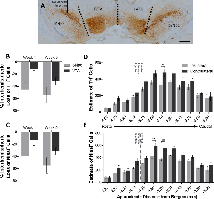
Parkinson's disease (PD) manifests clinically as bradykinesia, rigidity, and development of a resting tremor, primarily due to degeneration of dopaminergic nigrostriatal pathways in the brain. Intranigral administration of the irreversible ubiquitin proteasome system inhibitor, lactacystin, has been used extensively to model nigrostriatal degeneration in rats, and study the effects of candidate neuroprotective agents on the integrity of the dopaminergic nigrostriatal system. Recently however, adjacent extra-nigral brain regions such as the ventral tegmental area (VTA) have been noted to also become affected in this model, yet their integrity in studies of candidate neuroprotective agents in the model have largely been overlooked. Here we quantify the extent and distribution of dopaminergic degeneration in the VTA of rats intranigrally lesioned with lactacystin, and quantify the extent of VTA dopaminergic neuroprotection after systemic treatment with an epigenetic therapeutic agent, valproate, shown previously to protect dopaminergic SNpc neurons in this model. We found that unilateral intranigral administration of lactacystin resulted in a 53.81% and 31.72% interhemispheric loss of dopaminergic SNpc and VTA neurons, respectively. Daily systemic treatment of lactacystin lesioned rats with valproate however resulted in dose-dependant neuroprotection of VTA neurons. Our findings demonstrate that not only is the VTA also affected in the intranigral lactacystin rat model of PD, but that this extra-nigral brain region is substrate for neuroprotection by valproate, an agent shown previously to induce neuroprotection and neurorestoration of SNpc dopaminergic neurons in this model. Our results therefore suggest that valproate is a candidate for extra-nigral as well as intra-nigral neuroprotection.
Keywords: Dopamine; Histone deacetylase inhibitor; Lactacystin; Parkinson’s disease; Substantia Nigra pars compacta; Valproate; Ventral tegmental area.
Copyright © 2015 The Authors. Published by Elsevier Ireland Ltd.. All rights reserved.
Figures


References
-
- Braak H., Tredici K.D., Rüb U., de Vos R.A.I., Jansen Steur E.N.H., Braak E. Staging of brain pathology related to sporadic Parkinson’s disease. Neurobiol. Aging. 2003;24:197–211. - PubMed
-
- Broide R.S., Redwine J.M., Aftahi N., Young W., Bloom F.E., Winrow C.J. Distribution of histone deacetylases 1–11 in the rat brain. J. Mol. Neurosci. 2007;31:47–58. - PubMed
-
- Carman L.S., Gage F.H., Shults C.W. Partial lesion of the substantia nigra: relation between extent of lesion and rotational behavior. Brain Res. 1991;553:275–283. - PubMed
-
- Elson J., Yates A., Pienaar I. Pedunculopontine cell loss and protein aggregation direct microglia activation in parkinsonian rats. Brain Struct. Funct. 2015:1–23. - PubMed
Publication types
MeSH terms
Substances
Grants and funding
LinkOut - more resources
Full Text Sources
Other Literature Sources
Miscellaneous
