. 2016 Mar 1:316:209-20.
doi: 10.1016/j.neuroscience.2015.12.044. Epub 2015 Dec 30.

Density of GFAP-immunoreactive astrocytes is decreased in left hippocampi in major depressive disorder

Affiliations

Density of GFAP-immunoreactive astrocytes is decreased in left hippocampi in major depressive disorder

J A Cobb et al. Neuroscience. .

Abstract

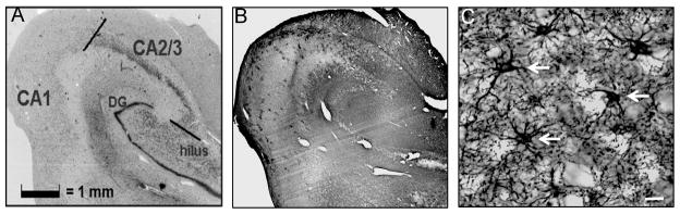
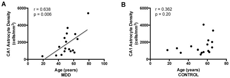
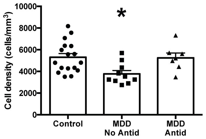
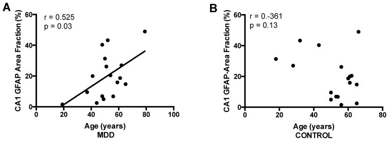
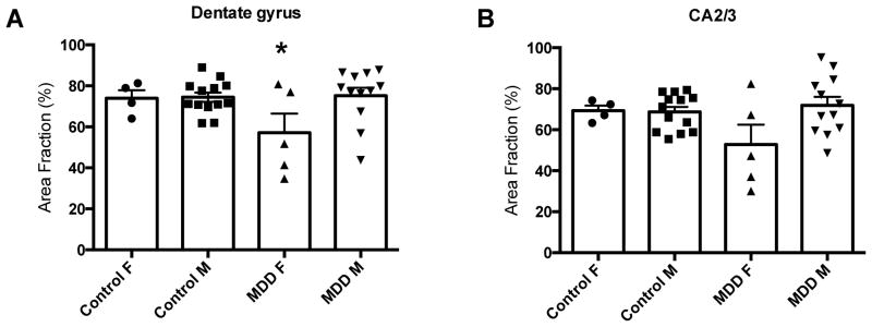
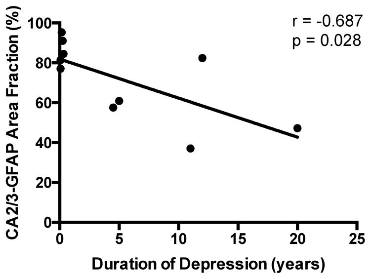
Neuroimaging and postmortem studies of subjects with major depressive disorder (MDD) reveal smaller hippocampal volume with lengthening duration of illness. Pathology in astrocytes may contribute significantly to this reduced volume and to the involvement of the hippocampus in MDD. Postmortem hippocampal tissues were collected from 17 subjects with MDD and 17 psychiatrically-normal control subjects. Sections from the body of the hippocampus were immunostained for glial fibrillary acidic protein (GFAP), a marker of intermediate filament protein expressed in astrocytes. The density of GFAP-immunoreactive astrocytes was measured in the hippocampus using 3-dimensional cell counting. Hippocampal subfields were also assessed for GFAP-immunoreactive area fraction. In CA1, there was a significant positive correlation between age and either density or area fraction in MDD. The density of astrocytes in the hilus, but not CA1 or CA2/3, was significantly decreased only in depressed subjects not taking an antidepressant drug, but not for depressed subjects taking an antidepressant drug. The area fraction of GFAP-immunoreactivity was significantly decreased in the dentate gyrus in women but not men with depression. In CA2/3, the area fraction of GFAP-immunoreactivity was inversely correlated with the duration of depression in suicide victims. Astrocyte contributions to neuronal function in the hilus may be compromised in depressed subjects not taking antidepressant medication. Due to the cross-sectional nature of the present study of postmortem brain tissue, it remains to be determined whether antidepressant drug treatment prevented a decrease in GFAP-immunoreactive astrocyte density or restored cell density to normal levels.

Keywords: GFAP; astrocyte; hippocampus; major depressive disorder; postmortem.


Figures

Figure 1
Figure 1
Photomicrographs of subregions of the human hippocampus. Nissl-stained section of the hippocampus (A) with adjacent GFAP-labeled section (B). High magnification image of GFAP-immunoreactive astrocytes in human hippocampus (C). Representative astrocytes noted by white arrows. Scale bar in C = 10 μm. Abbreviation: DG = dentate gyrus.
Figure 2
Figure 2
Correlation between age and GFAP-immunoreactive astrocyte density in the CA1 region of the hippocampus. There was a positive, significant correlation between astrocyte density and age in subjects with major depressive disorder (MDD) (A). There was no significant correlation between astrocyte density and age in control subjects (B).
Figure 3
Figure 3
GFAP-immunoreactive astrocyte density in the hilus. Astrocyte density was significantly decreased in subjects with major depressive disorder (MDD) with no antidepressant drug present postmortem (MDD, No Antid) compared to either control subjects or subjects with MDD with an antidepressant drug present postmortem (MDD, Antid). *ANCOVA, df = 2, F = 6.58, p = 0.005. p = 0.003 vs. control subjects and p = 0.011 vs. MDD, Antid. Histograms represent the mean ± S.E.M..
Figure 4
Figure 4
Correlation between age and GFAP-immunoreactive area fraction in the CA1 regions of the hippocampus. There was a positive, significant correlation between area fraction and age in subjects with major depressive disorder (MDD) (A) but not in control subjects (B).
Figure 5
Figure 5
GFAP-immunoreactive area fraction in control and major depressive disorder (MDD) men (M) and women (F). (A) After adjusting for age, there was a significant main effect of depression and gender in the dentate gyrus (ANCOVA, df = 3, F = 3.10, p = 0.043). * Females with MDD vs. males with MDD (p = 0.009), vs. male controls (p = 0.009), and a trend vs. female controls (p = 0.07). (B) After adjusting for age, there was a trend for a main effect of depression and gender in CA2/3 (ANCOVA, df = 3, F = 2.71, p = 0.064). Histograms represent the mean ± S.E.M..
Figure 6
Figure 6
Correlation between duration of MDD in suicide victims and GFAP-immunoreactive area fraction in CA2/3 region of the hippocampus. Area fraction was significantly decreased as a function of duration of illness.

References

    1. Amaral D, Lavenex P. Hippocampal neuroanatomy. In: Andersen P, Morris R, Amaral D, Bliss T, O’Keefe J, editors. The hippocampus book. New York: Oxford University Press; 2007. pp. 37–114.
    1. American Psychiatric Association (APA) Diagnostic and statistical manual of mental disorders. 4. Washington, DC: 1994.
    1. Araya-Callís C, Hiemke C, Abumaria N, Flugge G. Chronic psychosocial stress and citalopram modulate the expression of the glial proteins GFAP and NDRG2 in the hippocampus. Psychopharmacology. 2012;224:209–222. - PMC - PubMed
    1. Bachis A, Mallei A, Cruz MI, Wellstein A, Mocchetti I. Chronic antidepressant treatments increase basic fibroblast growth factor and fibroblast growth factor-binding protein in neurons. Neuropharmacology. 2008;55:1114–1120. - PMC - PubMed
    1. Banasr M, Duman RS. Glial loss in the prefrontal cortex is sufficient to induce depressive-like behaviors. Biol Psychiatry. 2008;64:863–870. - PMC - PubMed

Publication types

Substances