. 2016 Jan 8:6:19111.
doi: 10.1038/srep19111.

Enhanced engraftment, proliferation, and therapeutic potential in heart using optimized human iPSC-derived cardiomyocytes

Affiliations

Enhanced engraftment, proliferation, and therapeutic potential in heart using optimized human iPSC-derived cardiomyocytes

Shunsuke Funakoshi et al. Sci Rep. .

Abstract

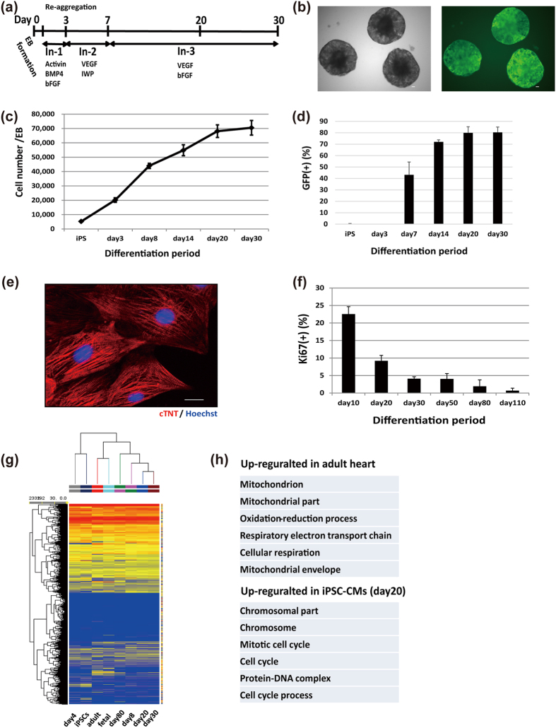
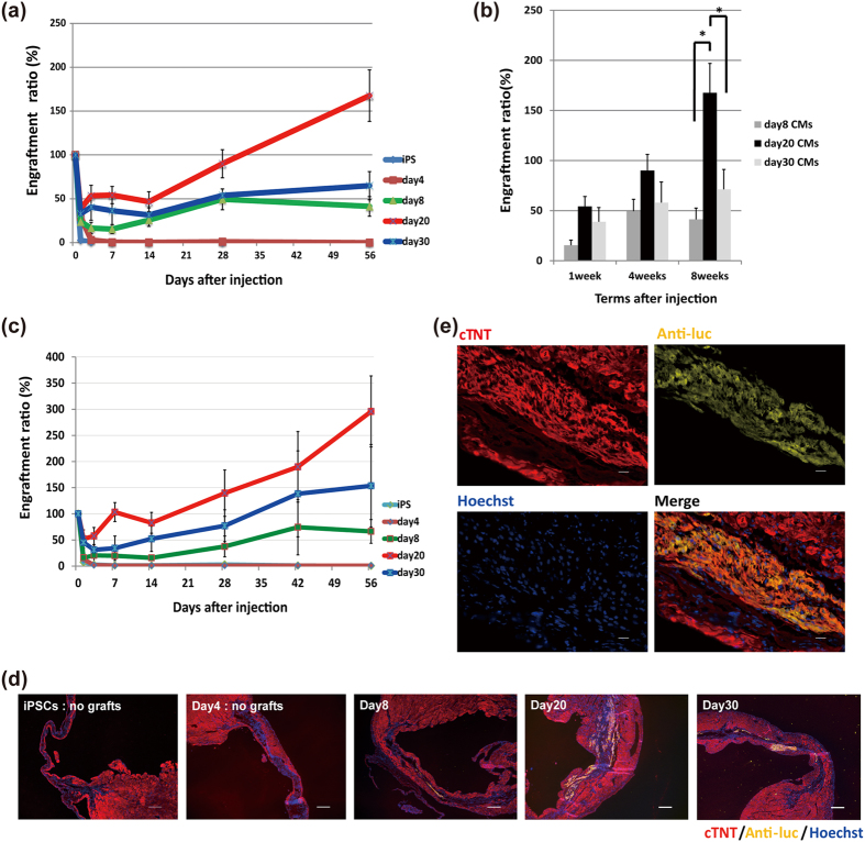
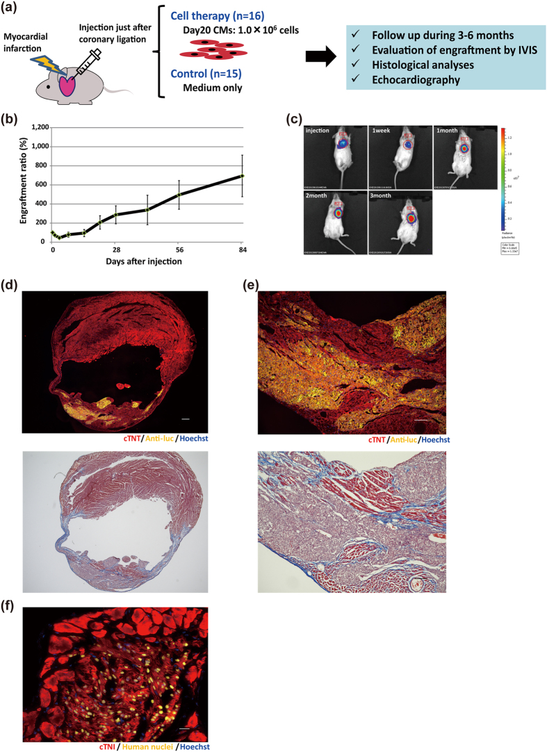
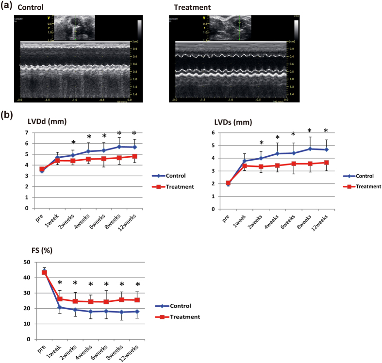
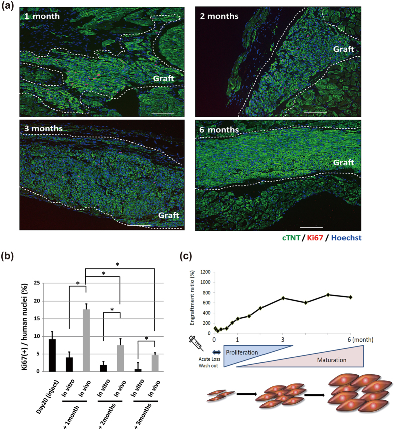
Human pluripotent stem cell-derived cardiomyocytes (CMs) are a promising tool for cardiac cell therapy. Although transplantation of induced pluripotent stem cell (iPSC)-derived CMs have been reported in several animal models, the treatment effect was limited, probably due to poor optimization of the injected cells. To optimize graft cells for cardiac reconstruction, we compared the engraftment efficiency of intramyocardially-injected undifferentiated-iPSCs, day 4 mesodermal cells, and day 8, day 20, and day 30 purified iPSC-CMs after initial differentiation by tracing the engraftment ratio (ER) using in vivo bioluminescence imaging. This analysis revealed the ER of day 20 CMs was significantly higher compared to other cells. Transplantation of day 20 CMs into the infarcted hearts of immunodeficient mice showed good engraftment, and echocardiography showed significant functional improvement by cell therapy. Moreover, the imaging signal and ratio of Ki67-positive CMs at 3 months post injection indicated engrafted CMs proliferated in the host heart. Although this graft growth reached a plateau at 3 months, histological analysis confirmed progressive maturation from 3 to 6 months. These results suggested that day 20 CMs had very high engraftment, proliferation, and therapeutic potential in host mouse hearts. They also demonstrate this model can be used to track the fate of transplanted cells over a long time.


Conflict of interest statement

S.Y. is a scientific advisor of iPS Academia Japan without salary.

Figures

Figure 1
Figure 1. Cardiac differentiation and characteristics of iPSC-derived cardiomyocytes.
(a) Schematic representation of the cardiac differentiation protocol. (b) Representative images of differentiated embryoid bodies. Left: bright-field; right: GFP. Scale bars: 100 μm. (c) Increasing number of cells per embryoid body during cardiac differentiation (n = 3). (d) Percentage of GFP-positive cells during differentiation (n = 3).(e) Immunostaining on day 20 of purified CMs: red, cTNT; blue, Hoechst. Scale bar: 20 μm. (f) Percentage of Ki67-positive CMs in vitro (n = 3) over different days. Data are represented as mean ± SD. (g) Hierarchical clustering of the global gene expression data obtained from undifferentiated iPSCs, day4 mesodermal cells, adult and fetal human hearts, and purified day8, 20, 30, and 80 iPSC-CMs. (h) Gene ontology analysis of differentially expressed genes between day20 iPSC-CMs and adult heart (fold change > 2.0, p < 0.05).
Figure 2
Figure 2. Engraftment capacity during different cardiac differentiation stages.
(a) Engraftment ratio in the first 2 months after injection into healthy heart. iPSCs and day4 mesodermal cells, n = 5 each; day8, 20, and 30 iPSC-CMs, n = 7 each. Values are mean ± SE. (b) Engraftment ratio of purified CMs (day8, 20, and 30, n = 7 each) at 1, 4, and 8 weeks after the initial injection. Values are mean ± SE. *p < 0.05 by one-way ANOVA followed by Tukey’s posthoc test. (c) Engraftment ratio in the first 2 months after injection into infarcted heart. iPSCs, day4 mesodermal cells, and day8, 20, and 30 iPSC-CMs, n = 4 each. Values are mean ± SE. (d) Immunostaining of engrafted CMs in infarcted heart at each differentiation stage for cTNT (red), anti-luciferase (luc) (yellow), and Hoechst (blue). Scale bars: 300 μm. (e) Immunostaining of engrafted CMs in infarcted hearts at high magnification: red, cTNT; yellow, anti-luc; blue, Hoechst. Scale bars: 20 μm.
Figure 3
Figure 3. Engraftment of iPSC-CMs in infarcted mouse hearts.
(a) Schematic summary of cell therapy using day20 CMs. (b) Engraftment ratio after injection of day20 iPSC-CMs based on luminescence imaging data (n = 16). Values are mean ± SE. (c) Representative images of luminescence signals after injection into the hearts of NOG mice with myocardial infarction. (d) Upper: Immunostaining of engrafted CMs in the whole heart for cTNT (red), anti-luc (yellow), and Hoechst (blue). Scale bar: 300 μm. Images were photographed by a fluorescence microscope, BZ-X700 (Keyence). Lower: Trichrome-staining of the additional section of the upper image. (e) Upper: Magnified image of (d). Red, cTNT; yellow, anti-luc; blue, Hoechst. Scale bar: 100 μm. Lower: Trichrome-staining of the additional section of the upper image. (f) Immunostaining of engrafted CMs at high magnification. Red, cTNI; yellow, human nuclei; blue, Hoechst. Scale bar: 20 μm.
Figure 4
Figure 4. Therapeutic effect of engrafted iPSC-CMs.
(a) Echocardiographic findings at 3 months. Left: control mouse; right: cell-treated mouse. Dotted lines: boundary of anterior and posterior walls of left ventricle. (b) Echocardiographic parameters (LVDd, LVDs, and FS). Values are mean ± SD. *p < 0.05 by the unpaired t test.
Figure 5
Figure 5. Follow-up analysis showed efficient engraftment over a long time and maturation of engrafted CMs in vivo in infarcted hearts.
(a) Engraftment ratio over 6 months after injection of day20 CMs based on luminescence imaging data (day0–3 months: n = 16; 3 months-6 months: n = 5). Values are mean ± SE. (b) The luminescence signal ratio at several time points. The ratio was defined as the luminescence signal intensity at the end of the time interval divided by the intensity at the beginning of the interval. Values are mean ± SE.(c) Immunostaining of engrafted CMs in the whole heart at 6 months: red, cTNT; yellow, anti-luc; blue, Hoechst. Scale bar: 300 μm. Images were photographed by a fluorescence microscope BZ-X700 (Keyence). (d) Magnified image of (c): red, cTNT; yellow, anti-luc; blue, Hoechst. Scale bar: 100 μm. (e) Immunostaining of engrafted CMs at high magnification: red, cTNI; yellow, human nuclei; blue, Hoechst. Scale bar: 20 μm. (f) Transmission electron micrographs of iPSC-CMs at 2 months and 6 months after transplantation. Z, Z-disk; M, M-band; I, I-band; H, H-band; A, A-band; Mit, Mitochondrion; FA, Fascia Adherens; Ds, Desmosome; Gap, Gap junction. Scale bars: 500 nm.
Figure 6
Figure 6. Proliferation capacity of iPSC-CMs in vivo in infarcted mouse hearts.
(a) Comparison of Ki67 immunostaining of engrafted CMs at several time points: green, cTNT; red, Ki67; blue, Hoechst. Grafted areas are enclosed by white dotted lines. Scale bars: 100 μm. (b) Percentage of Ki67-positive CMs at several time points. Values are mean ± SD. *p < 0.05 by one-way ANOVA followed by Tukey’s posthoc test. (c) Schematic summary of the luminescence signal time course shown in Fig. 5a after direct injection of day20 CMs.
Figure 7
Figure 7. Optimized cell transplantation using another iPSC line purified by cell sorting for the cardiac surface marker, SIRPA.
(a) Flow cytometry analysis 20 days after initial differentiation. SIPRA-positive and CD31, 49a, 90, 140b, and TRA1-60-negative cells were sorted. (b) Flow cytometry analysis of the sorted cells stained by cTNT. (c) Follow-up bioluminescence imaging of injected day20 purified-CMs. Values are mean ± SE. (d) Immunostaining of engrafted CMs 6 months after the initial injection of day20 CMs for cTNT (red), anti-luc (green), and Hoechst (blue). Scale bars: 300 μm. (e) Immunostaining of engrafted CMs at high magnification: red, actinin; yellow, anti-luc; blue, Hoechst. Scale bars: 20 μm.

References

    1. Stehlik J. et al. The Registry of the International Society for Heart and Lung Transplantation: Twenty-eighth Adult Heart Transplant Report–2011. The Journal of heart and lung transplantation: the official publication of the International Society for Heart Transplantation 30, 1078–1094 (2011). - PubMed
    1. Ptaszek L. M., Mansour M., Ruskin J. N. & Chien K. R. Towards regenerative therapy for cardiac disease. Lancet 379, 933–942 (2012). - PubMed
    1. Malliaras K. et al. Intracoronary cardiosphere-derived cells after myocardial infarction: evidence of therapeutic regeneration in the final 1-year results of the CADUCEUS trial (CArdiosphere-Derived aUtologous stem CElls to reverse ventricUlar dySfunction). Journal of the American College of Cardiology 63, 110–122 (2014). - PMC - PubMed
    1. Schachinger V. et al. Intracoronary bone marrow-derived progenitor cells in acute myocardial infarction. N Engl J Med 355, 1210–1221 (2006). - PubMed
    1. Tendera M. et al. Intracoronary infusion of bone marrow-derived selected CD34+CXCR4+ cells and non-selected mononuclear cells in patients with acute STEMI and reduced left ventricular ejection fraction: results of randomized, multicentre Myocardial Regeneration by Intracoronary Infusion of Selected Population of Stem Cells in Acute Myocardial Infarction (REGENT) Trial. Eur Heart J 30, 1313–1321 (2009). - PubMed

Publication types

MeSH terms