Observational Study
. 2016 Jan 8:20:3.
doi: 10.1186/s13054-015-1161-9.

Influence of different electrode belt positions on electrical impedance tomography imaging of regional ventilation: a prospective observational study

Affiliations
Observational Study

Influence of different electrode belt positions on electrical impedance tomography imaging of regional ventilation: a prospective observational study

Jan Karsten et al. Crit Care. .

Abstract

Background: Electrical impedance tomography (EIT) is a non-invasive bedside tool which allows an individualized ventilator strategy by monitoring tidal ventilation and lung aeration. EIT can be performed at different cranio-caudal thoracic levels, but data are missing about the optimal belt position. The main goal of this prospective observational study was to evaluate the impact of different electrode layers on tidal impedance variation in relation to global volume changes in order to propose a proper belt position for EIT measurements.

Methods: EIT measurements were performed in 15 mechanically ventilated intensive care patients with the electrode belt at different thoracic layers (L1-L7). All respiratory and hemodynamic parameters were recorded. Blood gas analyses were obtained once at the beginning of EIT examination. Off-line tidal impedance variation/tidal volume (TV/VT) ratio was calculated, and specific patterns of impedance distribution due to automatic and user-defined adjustment of the colour scale for EIT images were identified.

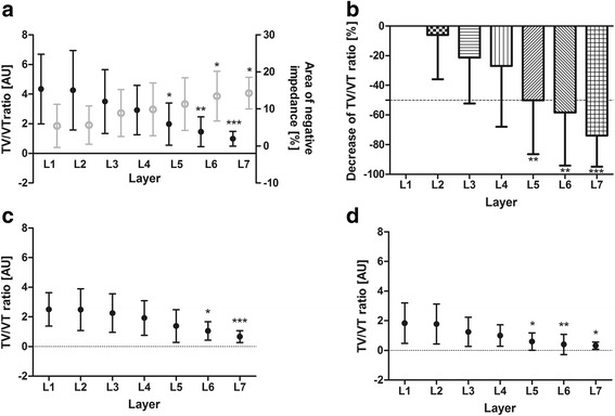
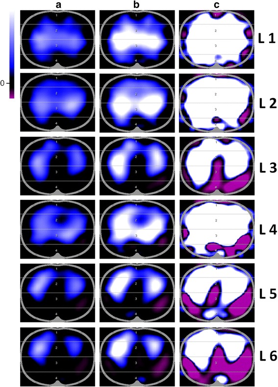
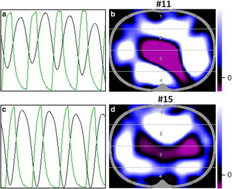
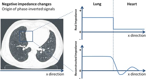
Results: TV/VT ratio is the highest at L1. It decreases in caudal direction. At L5, the decrease of TV/VT ratio is significant. We could identify patterns of diaphragmatic interference with ventilation-related impedance changes, which owing to the automatically adjusted colour scales are not obvious in the regularly displayed EIT images.

Conclusions: The clinical usability and plausibility of EIT measurements depend on proper belt position, proper impedance visualisation, correct analysis and data interpretation. When EIT is used to estimate global parameters like VT or changes in end-expiratory lung volume, the best electrode plane is between the 4th and 5th intercostal space. The specific colour coding occasionally suppresses user-relevant information, and manual rescaling of images is necessary to visualise this information.


Figures

Fig. 1
Fig. 1
TV/VT ratio at different layers. Interrelation of global TV/VT ratio (black) and amount of negative impedances (grey) (a). Percentage decrease of TV/VT ratio (relative) deeming L1 as baseline (b). Regional TV/VT ratio (c, d) at different layers from cranial (L1) to caudal (L7). c Non-dependent/ventral. d Dependent/dorsal. Data are mean ± standard deviation. Kruskal-Wallis test with Dunn’s post hoc testing for pairwise multiple comparison. Significance compared with L1: *P < 0.05, **P < 0.01, ***P < 0.001. AU arbitrary units, L layer, TV/VT tidal impedance variation/tidal volume ratio
Fig. 2
Fig. 2
Influence of belt position on electrical impedance tomography in patient 10. a TV/VT ratio depends on belt position. The ratio decreased in cranio-caudal direction (L1/ICS 4; L7/ICS 8). b Minute images of different layers in cranio-caudal direction were displayed as examples. Functional status images with automatically adjusted colour scales (assigning white to the maximum impedance change) are displayed on the left side. On the right side, colour scales were unadjusted to visualize interference. Images are a 32 × 32 pixel coloured matrix (white: positive/highest ∆Z; blue: medium ∆Z; black: impedance changes less than 10 %; purple: negative ∆Z, inverted signal). ICS intercostal space, L layer, TV/VT tidal impedance variation/tidal volume ratio
Fig. 3
Fig. 3
Influence of colour scales on functional electrical impedance tomography images. Examples of minute images in cranio-caudal direction (L1 to L6) (patient 1) with different colour scale endpoints: (a) automatically adjusted colour scales (assigning white to the maximum impedance change), (b, c) user-defined adjusted colour scales. Massive pleural effusion could be excluded. Images are 32 × 32 pixel coloured matrix (white: positive/highest ∆Z; blue: medium ∆Z; black: no impedance change; purple: negative ∆Z, inverted signal). L layer
Fig. 4
Fig. 4
Out-of-phase of impedance and tidal volume signal. a, c Impedance curves are painted black, and tidal volume curves are painted green. b, d Corresponding functional status images: the global impedance signals (blue/white) are induced mainly by the diaphragm (patient 11 L5, 12 cm below the armpits; patient 15 L7, 16 cm below the armpits)
Fig. 5
Fig. 5
Negative impedance changes. Origin of phase-inverted signals in reconstructed electrical impedance tomography images (representative example)

References

    1. Ahmed AH, Litell JM, Malinchoc M, Kashyap R, Schiller HJ, Pannu SR, et al. The role of potentially preventable hospital exposures in the development of acute respiratory distress syndrome: a population-based study. Crit Care Med. 2014;42:31–9. doi: 10.1097/CCM.0b013e318298a6db. - DOI - PMC - PubMed
    1. Futier E, Constantin JM, Paugam-Burtz C, Pascal J, Eurin M, Neuschwander A, et al. A trial of intraoperative low-tidal-volume ventilation in abdominal surgery. N Engl J Med. 2013;369:428–37. doi: 10.1056/NEJMoa1301082. - DOI - PubMed
    1. Severgnini P, Selmo G, Lanza C, Chiesa A, Frigerio A, Bacuzzi A, et al. Protective mechanical ventilation during general anesthesia for open abdominal surgery improves postoperative pulmonary function. Anesthesiology. 2013;118:1307–21. doi: 10.1097/ALN.0b013e31829102de. - DOI - PubMed
    1. Levin MA, McCormick PJ, Lin HM, Hosseinian L, Fischer GW. Low intraoperative tidal volume ventilation with minimal PEEP is associated with increased mortality. Br J Anaesth. 2014;113:97–108. doi: 10.1093/bja/aeu054. - DOI - PMC - PubMed
    1. Frerichs I, Becher T, Weiler N. Electrical impedance tomography imaging of the cardiopulmonary system. Curr Opin Crit Care. 2014;20:323–32. doi: 10.1097/MCC.0000000000000088. - DOI - PubMed

Publication types

LinkOut - more resources