. 2016 Jan 8:6:18969.
doi: 10.1038/srep18969.

Structural brain alterations in primary open angle glaucoma: a 3T MRI study

Affiliations

Structural brain alterations in primary open angle glaucoma: a 3T MRI study

Jieqiong Wang et al. Sci Rep. .

Abstract

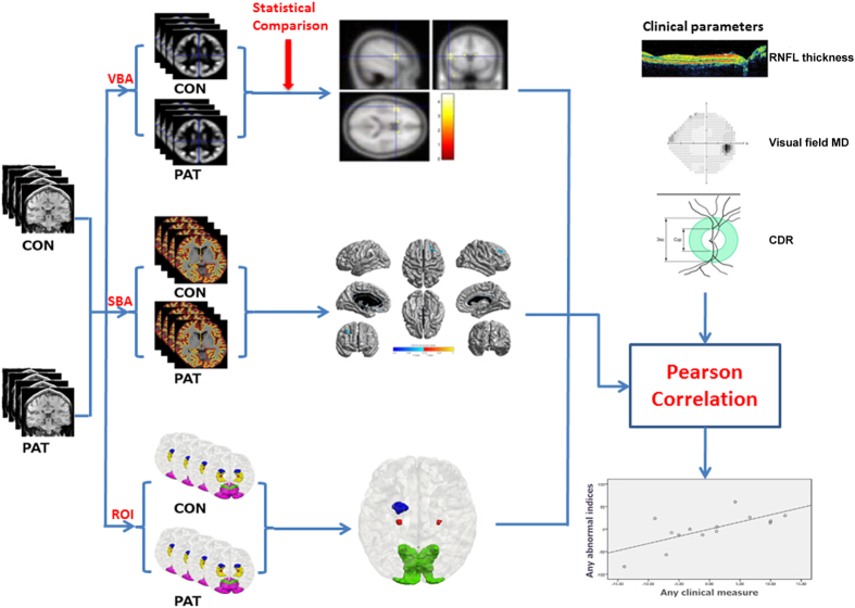
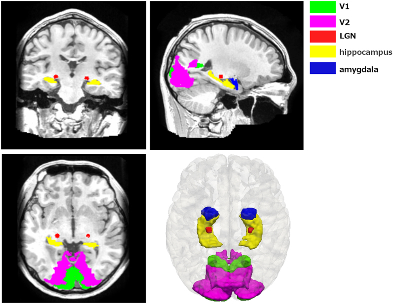
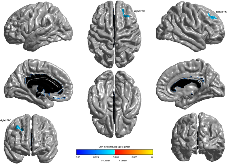
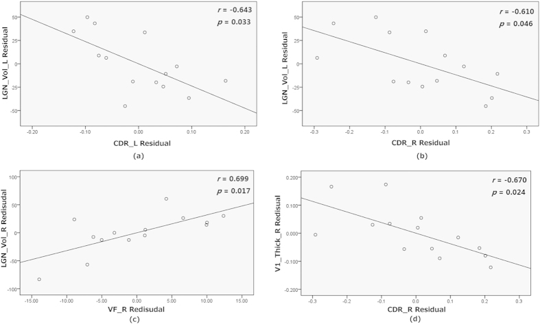
Glaucoma is not only an eye disease but is also associated with degeneration of brain structures. We now investigated the pattern of visual and non-visual brain structural changes in 25 primary open angle glaucoma (POAG) patients and 25 age-gender-matched normal controls using T1-weighted imaging. MRI images were subjected to volume-based analysis (VBA) and surface-based analysis (SBA) in the whole brain as well as ROI-based analysis of the lateral geniculate nucleus (LGN), visual cortex (V1/2), amygdala and hippocampus. While VBA showed no significant differences in the gray matter volumes of patients, SBA revealed significantly reduced cortical thickness in the right frontal pole and ROI-based analysis volume shrinkage in LGN bilaterally, right V1 and left amygdala. Structural abnormalities were correlated with clinical parameters in a subset of the patients revealing that the left LGN volume was negatively correlated with bilateral cup-to-disk ratio (CDR), the right LGN volume was positively correlated with the mean deviation of the right visual hemifield, and the right V1 cortical thickness was negatively correlated with the right CDR in glaucoma. These results demonstrate that POAG affects both vision-related structures and non-visual cortical regions. Moreover, alterations of the brain visual structures reflect the clinical severity of glaucoma.


Figures

Figure 1
Figure 1. The pipeline of the structural brain abnormality in glaucoma patients.
CON: normal controls, PAT: glaucoma patients, VBA: voxel-based analysis, SBA: surface-based analysis, ROI: ROI-based analysis, RNFL: retinal nerve fiber layer, CDR: cup-to-disk ratio, MD: mean deviation.
Figure 2
Figure 2. The visualization of structures in the ROI analysis.
Figure 3
Figure 3. Cortical regions with significantly reduced cortical thickness in glaucoma patients when compared with normal controls (P<0.05, FWE corrected). FPC: frontal pole cortex.
Figure 4
Figure 4. The partial correlation between the corresponding value of abnormal regions and clinical parameters with age, gender and intracerebral volume as covariates.
LGN_Vol_L/R: left/right LGN volume, V1_Thick_R: right V1 cortical thickness, CDR_L/R: left/right cup to disk ratio, VF_R: right visual field mean deviation. M Residue: the difference between M (the observed value) and the result (the theoretical value) of linear regression of M with age, gender and intracerebral volume.

References

    1. Guedes V. et al.. Optical coherence tomography measurement of macular and nerve fiber layer thickness in normal and glaucomatous human eyes. Ophthalmology 110, 177–189, doi: 10.1016/S0161-6420(02)01564-6 (2003). - DOI - PMC - PubMed
    1. Zhang Y. Q. et al.. Anterior visual pathway assessment by magnetic resonance imaging in normal-pressure glaucoma. Acta ophthalmologica 90, e295–302, doi: 10.1111/j.1755-3768.2011.02346.x (2012). - DOI - PubMed
    1. Chen W. W. et al.. Structural brain abnormalities in patients with primary open-angle glaucoma: a study with 3T MR imaging. Investigative ophthalmology & visual science 54, 545–554, doi: 10.1167/iovs.12-9893 (2013). - DOI - PubMed
    1. Greenfield D. S., Bagga H. & Knighton R. W. Macular thickness changes in glaucomatous optic neuropathy detected using optical coherence tomography. Arch Ophthalmol-Chic 121, 41–46 (2003). - PubMed
    1. Medeiros F. A. et al.. Evaluation of retinal nerve fiber layer, optic nerve head, and macular thickness measurements for glaucoma detection using optical coherence tomography. Am J Ophthalmol 139, 44–55, doi: 10.1016/j.ajo.2004.08.069 (2005). - DOI - PubMed

Publication types

LinkOut - more resources