. 2016 Mar 1;25(5):951-63.
doi: 10.1093/hmg/ddv628. Epub 2016 Jan 6.

LRRK2 BAC transgenic rats develop progressive, L-DOPA-responsive motor impairment, and deficits in dopamine circuit function

Affiliations

LRRK2 BAC transgenic rats develop progressive, L-DOPA-responsive motor impairment, and deficits in dopamine circuit function

Max Sloan et al. Hum Mol Genet. .

Abstract

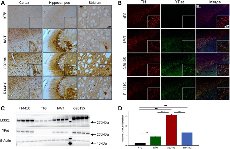
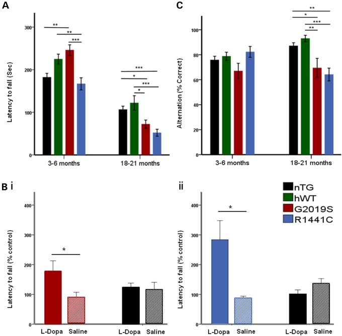
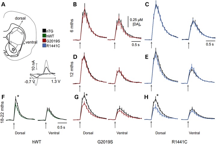
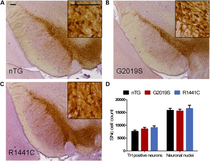
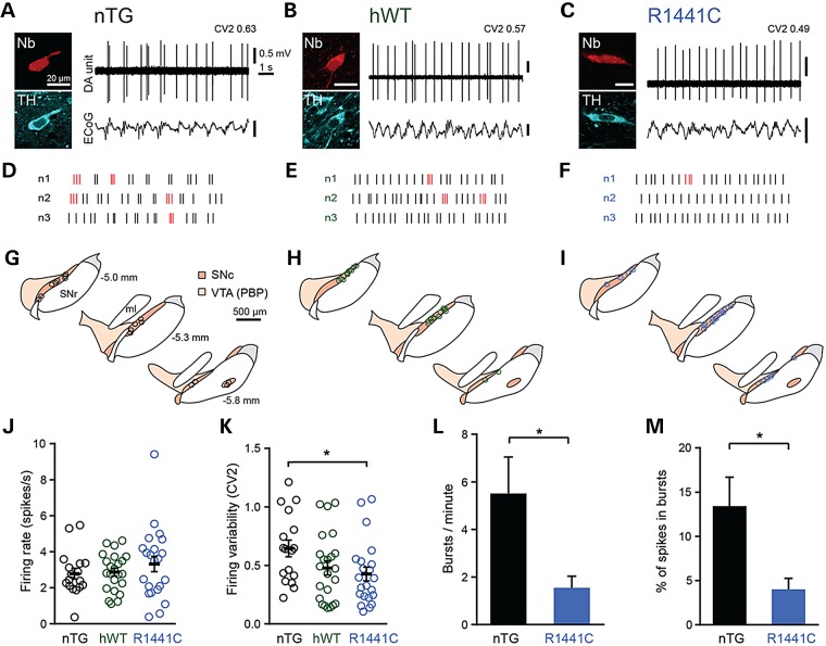
Mutations in leucine-rich repeat kinase 2 (LRRK2) lead to late-onset, autosomal dominant Parkinson's disease, characterized by the degeneration of dopamine neurons of the substantia nigra pars compacta, a deficit in dopamine neurotransmission and the development of motor and non-motor symptoms. The most prevalent Parkinson's disease LRRK2 mutations are located in the kinase (G2019S) and GTPase (R1441C) encoding domains of LRRK2. To better understand the sequence of events that lead to progressive neurophysiological deficits in vulnerable neurons and circuits in Parkinson's disease, we have generated LRRK2 bacterial artificial chromosome transgenic rats expressing either G2019S or R1441C mutant, or wild-type LRRK2, from the complete human LRRK2 genomic locus, including endogenous promoter and regulatory regions. Aged (18-21 months) G2019S and R1441C mutant transgenic rats exhibit L-DOPA-responsive motor dysfunction, impaired striatal dopamine release as determined by fast-scan cyclic voltammetry, and cognitive deficits. In addition, in vivo recordings of identified substantia nigra pars compacta dopamine neurons in R1441C LRRK2 transgenic rats reveal an age-dependent reduction in burst firing, which likely results in further reductions to striatal dopamine release. These alterations to dopamine circuit function occur in the absence of neurodegeneration or abnormal protein accumulation within the substantia nigra pars compacta, suggesting that nigrostriatal dopamine dysfunction precedes detectable protein aggregation and cell death in the development of Parkinson's disease. In conclusion, our longitudinal deep-phenotyping provides novel insights into how the genetic burden arising from human mutant LRRK2 manifests as early pathophysiological changes to dopamine circuit function and highlights a potential model for testing Parkinson's therapeutics.


Figures

Figure 1.
Figure 1.
Characterization of transgene expression patterns in the brains of LRRK2 transgenic rats. (A) Peroxidase-immunohistochemistry for YPet protein in cortical, hippocampal and striatal sections of 3-month-old animals from each of the transgenic lines (G2019S, R1441C, hWT) compared with an nTG control. (B) Double immunofluorescence labelling shows the co-localization of LRRK2-YPet fusion protein and TH in neurons of the SNc of 3-month-old animals from each of the transgenic lines (G2019S, R1441C, hWT) and a nTG control. (C) Whole-brain homogenate Western blots for YPet and LRRK2 in 3-month-old hWT, G2019S, R1441C and nTG rats. The molecular marker lane is run between hWT and G2019S lanes and the molecular weights of the reference bands are indicated (arrows). (D) Quantification of LRRK2 protein levels revealed overexpression of human LRRK2 was roughly 4–5× for hWT and R1441C, and 12× for G0291S compared with endogenous rat LRRK2 (One-way ANOVA: main effect of genotype: P < 0.0001; n = 4 per genotype). Bonferroni post hoc tests **P < 0.01, ***P < 0.001. Data are expressed as mean ± SEM.
Figure 2.
Figure 2.
Mutant LRRK2 transgenic rats develop late-stage l-DOPA-responsive motor deficits and cognitive impairment. (A) At 3–6 months G2019S, but not R1441C, animals performed better on the rotarod than nTG controls (age/genotype interaction: P < 0.0001). One-way ANOVA main effect of genotype, *P < 0.05, **P < 0.01, ***P < 0.001, n = 8 per, Tukey HSD post hoc test). At 18–21 months, rotarod performance was impaired in both G2019S and R1441C rats when compared with nTG and hWT controls (one-way ANOVA main effect of genotype, *P < 0.05, **P < 0.01, ***P < 0.001 n = 6–11 per genotype, Tukey HSD post hoc test. Rotarod data transformed (square-rooted to comply with parametric testing but presented as non-transformed data for ease of the reader). (B (i)) l-DOPA reversed the motor deficit in 18- to 21-month-old G2019S rats when compared with saline treatment (two-tailed Mann–Whitney U, P < 0.05, n = 6 per genotype). There was no significant difference in performance between l-DOPA-treated 18- to 21-month-old nTG rats and saline treated rats (two-tailed Mann–Whitney U, P > 0.05, n = 4 per genotype). (B ii) l-DOPA treatment of 21-month-old R1441C rats reversed the deficit in rotarod performance compared with saline controls (T-test, P < 0.05, n = 6 per genotype). Again no significant improvement in performance was seen in l-DOPA-treated nTG rats compared with saline controls (n = 5–6 per genotype). (C) Spontaneous alternation performance was impaired in old but not young LRRK2 G2019S and R1441C mutant rats compared with nTG and hWT controls (age/genotype interaction: P < 0.05). At 3–6 months, performance of G2019S and R1441C animals in the spontaneous alternation test was similar compared with nTG and hWT controls (one-way ANOVA main effect of genotype, *P < 0.05, n = 16 per genotype, Tukey HSD post hoc test). However, at 18–21 months, LRRK2 R1441C and G2019S rats showed significantly impaired performance on the spontaneous alternation test compared with nTG and hWT controls (one-way ANOVA main effect of genotype, *P < 0.05, **P < 0.01, ***P < 0.001, n = 9–14 per genotype, Tukey HSD post hoc test. Data are expressed as mean ± SEM.
Figure 3.
Figure 3.
LRRK2 transgenic rats develop late-stage dopamine transmission deficits in the dorsal striatum. (A) Schematic of recording site locations in striatal slices from R1441C, G2019S, hWT and nTG rats. Representative cyclic voltammogram (inset) recorded in striatal tissue (black line) shows peaks at the same oxidation and reduction potentials as a 2 µM dopamine calibration voltammogram (grey line). (B–H) Mean [DA]o profiles versus time following single pulse stimulation (↑200 µs, 600 µA) in dorsal (left panel) or ventral striatum (right panel) of (B, C) 6-month-old G2019S and R1441C rats, (D, E) 12-month-old G2019S and R1441C rats and (F–H) 18- to 22-month-old hWT, G2019S and R1441C rats. Mean peak evoked [DA]o was not significantly different between R1441C rats and nTG controls in either dorsal or ventral striatum at 6 and 12 months of age (P > 0.05; unpaired two-tailed Mann–Whitney; n = 15–29); however, at 18–21 months, mean peak evoked [DA]o was significantly lower in R1441C (0.30 ± 0.06 µM) compared with nTG rats (0.49 ± 0.06 µM) in dorsal striatum (*P < 0.05; unpaired two-tailed Mann–Whitney test; n = 15) but not ventral striatum (P > 0.05; unpaired two-tailed Mann–Whitney test; n = 12). Mean peak evoked [DA]o was not significantly different between G2019S rats and nTG controls in either dorsal or ventral striatum at 6 and 12 months of age (P > 0.05; unpaired two-tailed Mann–Whitney test; n = 11–21); however, at 18–21 months, mean peak evoked [DA]o was significantly lower in G2019S rats (0.40 ± 0.04 µM) compared with nTG controls (0.59 ± 0.07 µM) in dorsal striatum (*P < 0.05; unpaired two-tailed Mann–Whitney test; n = 36) but not ventral striatum (P > 0.05; unpaired two-tailed Mann–Whitney test; n = 25). At 22 months, mean peak evoked [DA]o was significantly lower in LRRK2 hWT rats (0.27 ± 0.03 µM) compared with nTG controls (0.37 ± 0.04 µM) in dorsal striatum (*P < 0.05; unpaired two-tailed Mann–Whitney test; n = 30) but not ventral striatum (P > 0.05; unpaired two-tailed Mann–Whitney test; n = 29–30). Data are expressed as mean ± SEM. Comparison of falling phases of mean dopamine release transients from CPu of 18- to 22-month-old nTG and LRRK2 transgenic rats reveals no significant difference (one-phase exponential decay curve fits; P > 0.05, n = 15–35, K = 2.94–2.97 s−1 nTG K = 2.82–3.19 s−1 LRRK2 transgenics), suggesting no difference in reuptake rate of dopamine between genotypes.
Figure 4.
Figure 4.
LRRK2 transgenic rats do not exhibit neurodegeneration in the SNc. (A–C) Sections of the LRRK2 transgenic SNc, immunolabelled for TH, that were used for stereological estimates. (D) Stereological estimates of the number of TH-positive neurons revealed no differences between the SNc of 18- to 21-month-old G2019S, R1441C and nTG rats. (One-way ANOVA: no main effect of genotype: P > 0.05, n = 5–10 per genotype). Similarly, there were no differences in the numbers neuronal nuclei populations between genotypes (one-way ANOVA: no main effect of genotype: P > 0.05, n = 5–10 per genotype). Scale bars, 200 µm.
Figure 5.
Figure 5.
In vivo firing pattern of SNc dopamine neurons is more regular in aged R1441C rats. Spontaneous activity of identified SNc dopamine neurons (DA unit) in 16- to 22-month-old nTG (A, D, G), hWT (B, E, H) and R1441C rats (C, F, I) during robust slow-wave activity (measured in the electrocorticogram, ECoG). After recording, individual neurons were juxtacellularly labelled with Neurobiotin and confirmed to be dopaminergic by expression of TH immunoreactivity. (DF) Example raster plots denoting 10 s of spike firing in three example neurons from each genotype. Spikes detected as occurring within bursts (see Supplementary Material, methods) are highlighted in red. (GI) Coronal schematics with approximate locations of recorded and labelled dopamine neurons for each genotype on three rostro-caudal levels (distance caudal to Bregma shown in G; dorsal top, lateral right). PBP, parabrachial pigmented area of the ventral tegmental area; SNc, substantia nigra pars compacta; SNr, substantia nigra pars reticulata; ml, medial lemniscus (adapted from (50)). (J,K) Mean firing rate and firing variability of SNc dopamine neurons (nTG n = 17 neurons, hWT n = 23 neurons, R1441C n = 22 neurons, *P < 0.05, one-way ANOVA on ranks, with Dunn's post hoc test). (L,M) Mean number of bursts per minute and mean percentage of spikes occurring within bursts (*P < 0.05, Mann–Whitney rank-sum test). Data in J–M are displayed as mean ± SEM. In (J) and (K), each circle represents data from a single neuron.

References

    1. Healy D.G., Falchi M., O'Sullivan S.S., Bonifati V., Durr A., Bressman S., Brice A., Aasly J., Zabetian C.P., Goldwurm S. et al. (2008) Phenotype, genotype, and worldwide genetic penetrance of LRRK2-associated Parkinson’s disease: a case–control study. Lancet Neurol., 7, 583–590. - PMC - PubMed
    1. Haugarvoll K., Wszolek Z.K. (2009) Clinical features of LRRK2 parkinsonism. Parkinsonism Relat. Disord., 15(Suppl. 3), S205–S208. - PubMed
    1. Cookson M.R. (2010) The role of leucine-rich repeat kinase 2 (LRRK2) in Parkinson’s disease. Nat. Rev. Neurosci., 11, 791–797. - PMC - PubMed
    1. Zimprich A., Biskup S., Leitner P., Lichtner P., Farrer M., Lincoln S., Kachergus J., Hulihan M., Uitti R.J., Calne D.B. et al. (2004) Mutations in LRRK2 cause autosomal-dominant parkinsonism with pleomorphic pathology. Neuron, 44, 601–607. - PubMed
    1. Gilks W.P., Abou-Sleiman P.M., Gandhi S., Jain S., Singleton A., Lees A.J., Shaw K., Bhatia K.P., Bonifati V., Quinn N.P. et al. (2005) A common LRRK2 mutation in idiopathic Parkinson's disease. Lancet, 365, 415–416. - PubMed

Publication types

MeSH terms

Substances

Supplementary concepts