. 2016 Apr;130(8):575-86.
doi: 10.1042/CS20150697. Epub 2016 Jan 7.

Epigenetic regulation of cyclooxygenase-2 by methylation of c8orf4 in pulmonary fibrosis

Affiliations

Epigenetic regulation of cyclooxygenase-2 by methylation of c8orf4 in pulmonary fibrosis

Iona C Evans et al. Clin Sci (Lond). 2016 Apr.

Abstract

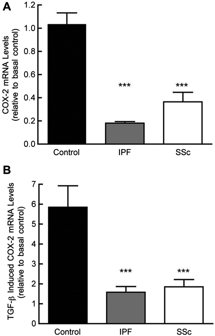
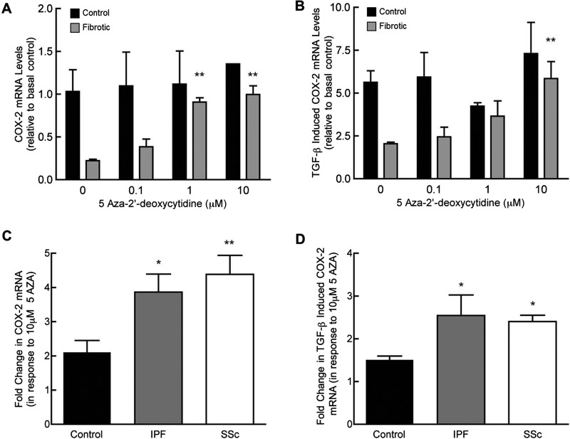
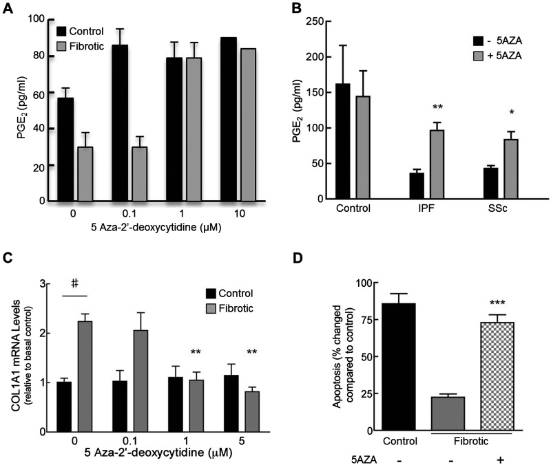
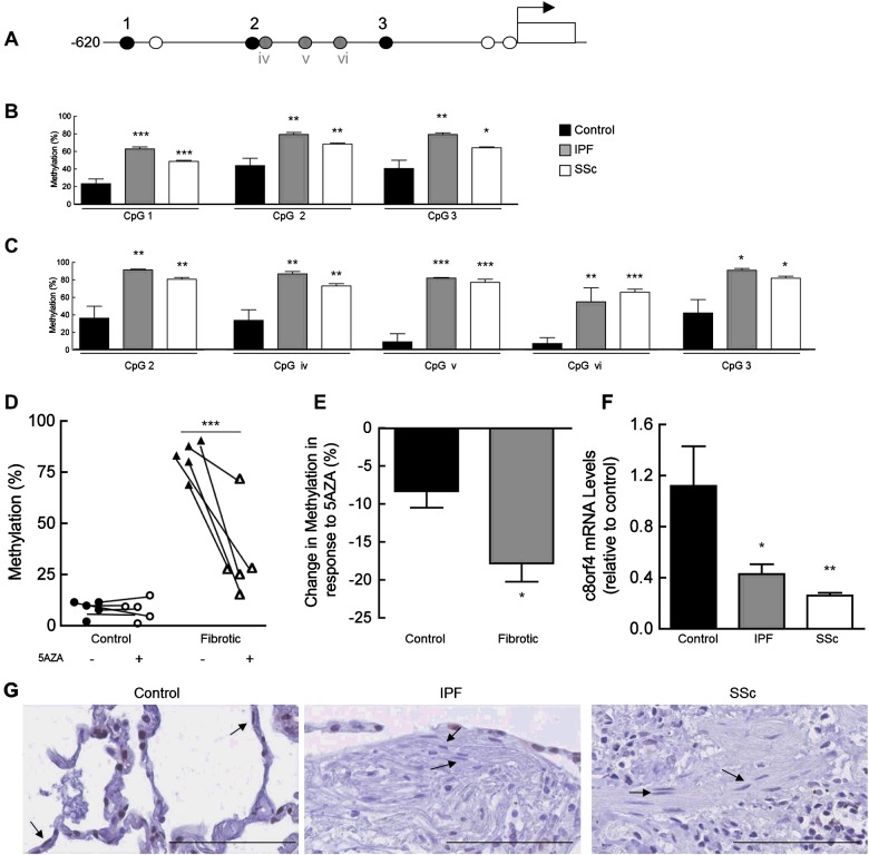
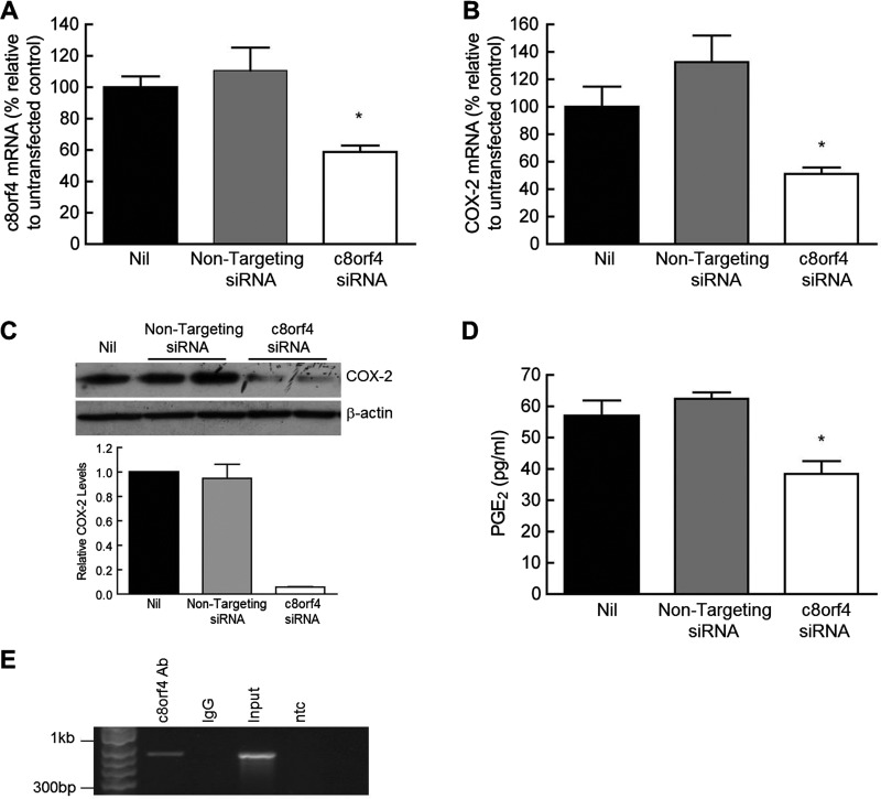
Fibroblasts derived from the lungs of patients with idiopathic pulmonary fibrosis (IPF) and systemic sclerosis (SSc) produce low levels of prostaglandin (PG) E2, due to a limited capacity to up-regulate cyclooxygenase-2 (COX-2). This deficiency contributes functionally to the fibroproliferative state, however the mechanisms responsible are incompletely understood. In the present study, we examined whether the reduced level of COX-2 mRNA expression observed in fibrotic lung fibroblasts is regulated epigenetically. The DNA methylation inhibitor, 5-aza-2'-deoxycytidine (5AZA) restored COX-2 mRNA expression by fibrotic lung fibroblasts dose dependently. Functionally, this resulted in normalization of fibroblast phenotype in terms of PGE2 production, collagen mRNA expression and sensitivity to apoptosis. COX-2 methylation assessed by bisulfite sequencing and methylation microarrays was not different in fibrotic fibroblasts compared with controls. However, further analysis of the methylation array data identified a transcriptional regulator, chromosome 8 open reading frame 4 (thyroid cancer protein 1, TC-1) (c8orf4), which is hypermethylated and down-regulated in fibrotic fibroblasts compared with controls. siRNA knockdown of c8orf4 in control fibroblasts down-regulated COX-2 and PGE2 production generating a phenotype similar to that observed in fibrotic lung fibroblasts. Chromatin immunoprecipitation demonstrated that c8orf4 regulates COX-2 expression in lung fibroblasts through binding of the proximal promoter. We conclude that the decreased capacity of fibrotic lung fibroblasts to up-regulate COX-2 expression and COX-2-derived PGE2 synthesis is due to an indirect epigenetic mechanism involving hypermethylation of the transcriptional regulator, c8orf4.

Keywords: DNA methylation; cyclooxygenase-2; fibroblast; idiopathic pulmonary fibrosis; prostaglandin E2; systemic sclerosis.


Figures

Figure 1
Figure 1. Basal and TGF-β-induced levels of COX-2 mRNA in control and fibrotic lung fibroblasts
Fibroblasts were grown to confluence, serum deprived for 24 h, and then incubated for 3 h in medium alone (A) or in TGF-β1 at a concentration of 1 ng/ml (B), before RNA was extracted for quantitative real-time PCR. Data are enumerated as mRNA levels relative to mean basal levels in control cell lines. Each bar represents the mean ± S.E.M. for four control, seven IPF and four SSc donor cell isolates. (***P<0.001 compared with control, one-way ANOVA with Tukey's post-test).
Figure 2
Figure 2. DNMT inhibition preferentially increases COX-2 mRNA levels in fibrotic lung fibroblasts
Proliferating cultures of control and fibrotic lung fibroblast lines were incubated with indicated concentrations of 5AZA for 96 h. Fibroblasts were then treated with medium alone (A) or with TGF-β1 (1 ng/ml) for 3 h (B) before being harvested for COX-2 quantitative real-time PCR. Data are enumerated as the fold increase, relative to mean mRNA levels basally in control cells. Each bar represents the mean of duplicate samples ± S.E.M. for one representative control and fibrotic cell line (**P<0.01 compared with 0 μM 5AZA, two-way ANOVA with Bonferroni post-test). Proliferating cultures were incubated with 10 μM of 5AZA for 96 h and then treated with medium alone (C) or with TGF-β1 (1 ng/ml) for 3 h (D) before being harvested for COX-2 quantitative real-time PCR. Data are enumerated as the fold increase, relative to mean levels in non-5AZA treated cells. Each bar represents mean ± S.E.M. for four control, seven IPF and four SSc donor cell isolates (*P<0.05, **P<0.01, compared with control group, one-way ANOVA with Tukey's post-test).
Figure 3
Figure 3. DNMT inhibition restores fibrotic lung fibroblast PGE2, collagen production and sensitivity to FasL-induced apoptosis
Proliferating cultures of single representative control and fibrotic lung fibroblast isolates were treated with 0, 0.1, 1 or 10 μM 5AZA for 96 h. Cells were serum deprived for 24 h before cell supernatants were harvested for PGE2 enzyme immunoassay. (A) Each bar represents the mean of duplicate samples ± S.E.M. (where no error bar is shown, only one sample was available for analysis). Cells from each group (control; n=4, IPF; n=7, SSc; n=4) were grown with 10 μM 5AZA before cell supernatants were collected for measuring PGE2 production (B). Each bar represents the mean ± S.E.M. (*P<0.05, **P<0.01, compared with control group, two-way ANOVA with Bonferroni post-test). Representative control and fibrotic lung fibroblasts were incubated with 5AZA before being harvested for collagen 1A qPCR (C). Data are enumerated as the fold increase, relative to mean mRNA levels basally in control cells. Each bar represents the mean ± S.E.M. for three independent experiments (**P<0.01 compared with 0 μM 5AZA, two-way ANOVA with Bonferroni post-test. #P<0.01 compared with basal levels in control). Representative control and fibrotic lung fibroblasts were grown in the presence of either 10 μM 5AZA or vehicle alone, in medium containing 10% FBS for 72 h (D). Fibroblasts were then serum starved overnight with or without 5AZA prior to being exposed to FasL (50 ng/ml) for 24 h. Apoptosis was determined by Annexin V/PI staining with FACS analysis. Bars represent the mean ± S.E.M. for six experimental replicates (***P<0.001 compared with non-5AZA treated, two-way ANOVA with Bonferroni post-test).
Figure 4
Figure 4. Increased methylation and decreased expression of c8orf4 in fibrotic lung fibroblasts
A schematic representation of the upstream 5′ region of c8orf4 (A). Circles indicate the location of CpG sites. Black circles represent the three CpG sites on the Illumina array, grey circles represent three additional sites tested by bisulfite sequencing and white circles represent untested CpG sites within the region. Genomic DNA extracted from primary fibroblasts was assessed and mean percentage methylation at each CpG site within each group is shown (B). Genomic DNA extracted from control, IPF and SSc cell lines were bisulfite treated and subjected to bisufite sequencing (C). Five CpG sites within the c8orf4 promoter were analysed. Mean percentage methylation at each CpG site within each of the groups is shown (C). For (B, C, E and F) data are derived from six control, five IPF and seven SSc donor fibroblast isolates. ***P<0.001, **P<0.01 and *P<0.05, compared with levels in control cells, one-way ANOVA with Tukey's post-test. DNA extracted from representative control and fibrotic fibroblasts treated with or without 1 μM 5AZA for ≥1 week was subjected to bisufite sequencing, as above (D). Each point represents the % methylation at the five individual CpG sites analysed in panel C (***P<0.001 compared with non-5Aza treated, two-way ANOVA with Bonferroni post-test). Control, IPF and SSc lung fibroblast lines were treated with 1 μM 5AZA as above, and DNA subjected to bisulfite sequencing (E). Data are enumerated as mean % DNA methylation across all five CpG sites within each group (*P<0.05, compared with levels in control cells). RNA extracted from control and fibrotic cell lines was subjected to qPCR for c8orf4 mRNA (F). Data are enumerated as mRNA levels relative to mean basal levels in control cells (±S.E.M., **P<0.01 and *P<0.05, one-way ANOVA with Tukey's post-test). Representative photomicrographs of control (n=3), IPF (n=6) and SSc (n=1) lung showing immunohistochemical localization of c8orf4 stained brown/black (G). Arrows indicate nuclear c8orf4 staining in control lung fibroblast-like cells but staining was weak or undetectable in fibrotic lung fibroblasts (scale bar represents 50 μm).
Figure 5
Figure 5. c8orf4 associates with the COX-2 promoter and knockdown in control fibroblasts down-regulates expression of COX-2 and PGE2 production
Control fibroblasts were transfected with either a non-targeting negative control or c8orf4 siRNA (10 nM) and RNA was extracted for qPCR (A and B). Levels of c8orf4 (A) and COX-2 (B) mRNA are represented as a percentage relative to non-transfected cells (Nil). Each bar represents mean ± S.E.M. for three independent experiments. (*P<0.05 compared with Nil, one-way ANOVA with Tukey's post-test). Protein samples were collected from cells transfected as above, and samples Western blotted for COX-2 (C). Relative COX-2 levels are expressed as a ratio of COX-2/β-actin and represented relative to levels in non-transfected cells. Cells were transfected as above, and cell supernatants were harvested for PGE2 enzyme immunoassay (D). Each bar represents the mean of triplicate wells ± S.E.M. and is representative of three individual experiments (*P<0.05 compared with non-transfected control, one-way ANOVA with Tukey's post-test). Chromatin extracted from representative primary control fibroblasts was immunoprecipitated with a c8orf4 antibody followed by PCR with a panel of COX-2 promoter primer sets (E). ChIP PCR products, corresponding to region −579 to −1271, were visualized by agarose gel electrophoresis (IgG corresponds to isotype control; input is sonicated but non-immunoprecipitated sample and ntc is no template control for the PCR).

Similar articles

Cited by

References

    1. Maher T.M., Wells A.U., Laurent G.J. Idiopathic pulmonary fibrosis: multiple causes and multiple mechanisms? Eur. Respir. J. 2007;30:835–839. doi: 10.1183/09031936.00069307. - DOI - PubMed
    1. Varga J., Abraham D. Systemic sclerosis: a prototypic multisystem fibrotic disorder. J. Clin. Invest. 2007;117:557–567. doi: 10.1172/JCI31139. - DOI - PMC - PubMed
    1. Gribbin J., Hubbard R.B., Le Jeune I., Smith C.J., West J., Tata L.J. Incidence and mortality of idiopathic pulmonary fibrosis and sarcoidosis in the UK. Thorax. 2006;61:980–985. doi: 10.1136/thx.2006.062836. - DOI - PMC - PubMed
    1. McAnulty R.J. Fibroblasts and myofibroblasts: their source, function and role in disease. Int. J. Biochem. Cell Biol. 2007;39:666–671. doi: 10.1016/j.biocel.2006.11.005. - DOI - PubMed
    1. Rosenbloom J., Mendoza F.A., Jimenez S.A. Strategies for anti-fibrotic therapies. Biochim. Biophys. Acta. 2013;1832:1088–1103. doi: 10.1016/j.bbadis.2012.12.007. - DOI - PubMed

Publication types

MeSH terms