

Human Cortical Neural Stem Cells Expressing Insulin-Like Growth Factor-I: A Novel Cellular Therapy for Alzheimer's Disease
- PMID: 26744412
- PMCID: PMC4807660
- DOI: 10.5966/sctm.2015-0103
Human Cortical Neural Stem Cells Expressing Insulin-Like Growth Factor-I: A Novel Cellular Therapy for Alzheimer's Disease
Abstract
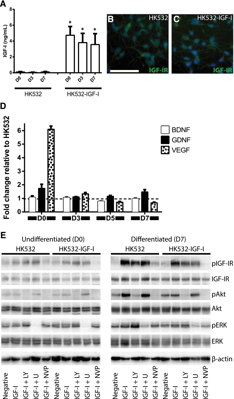
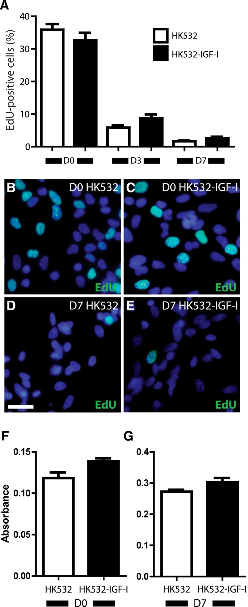
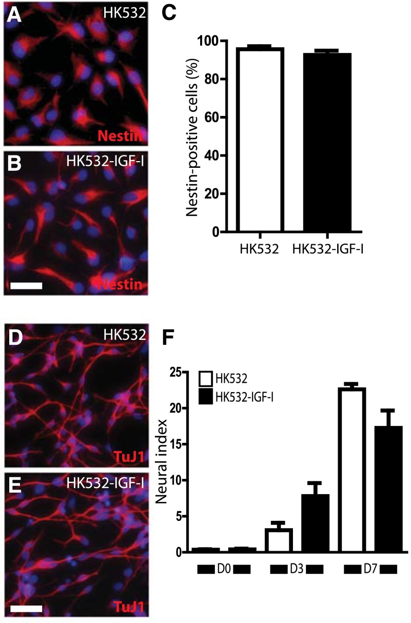
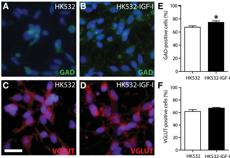
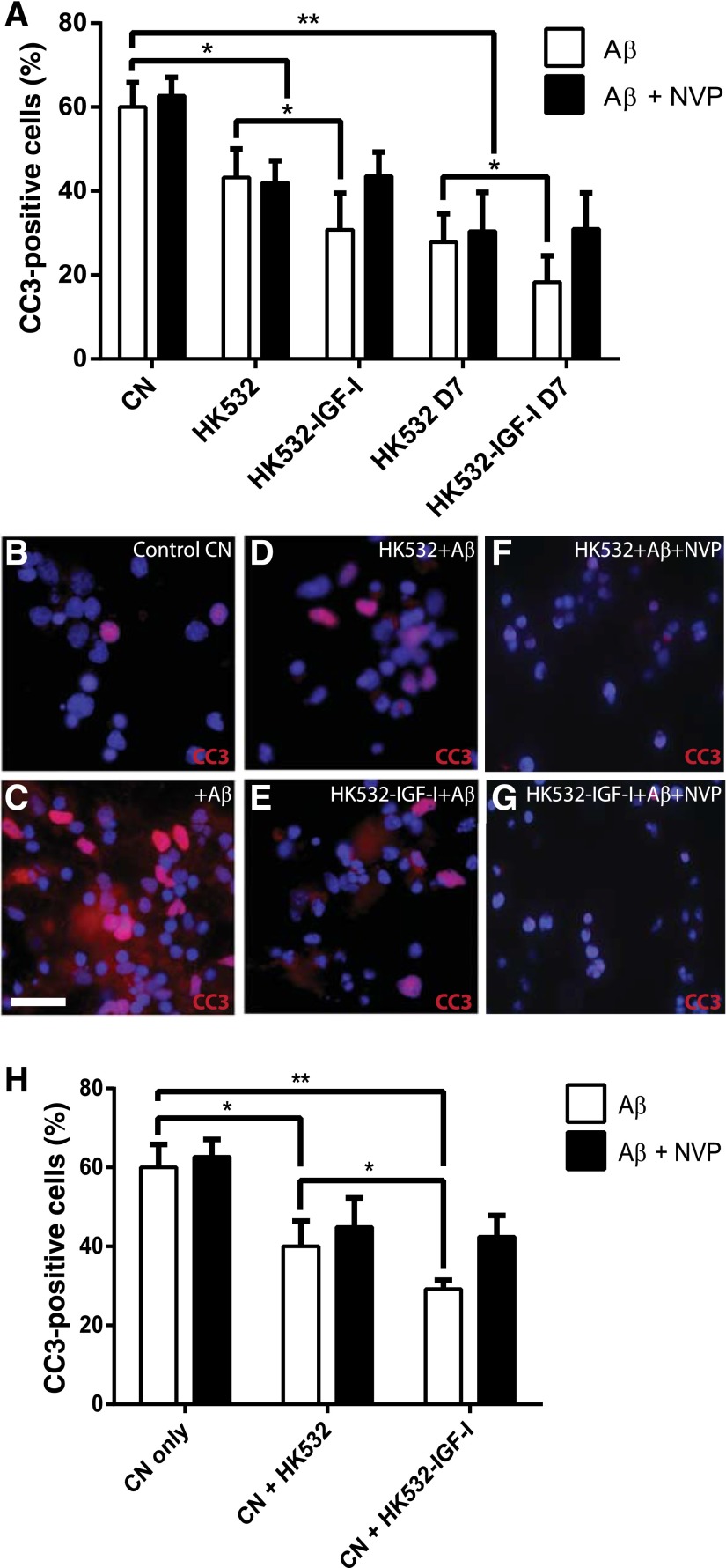
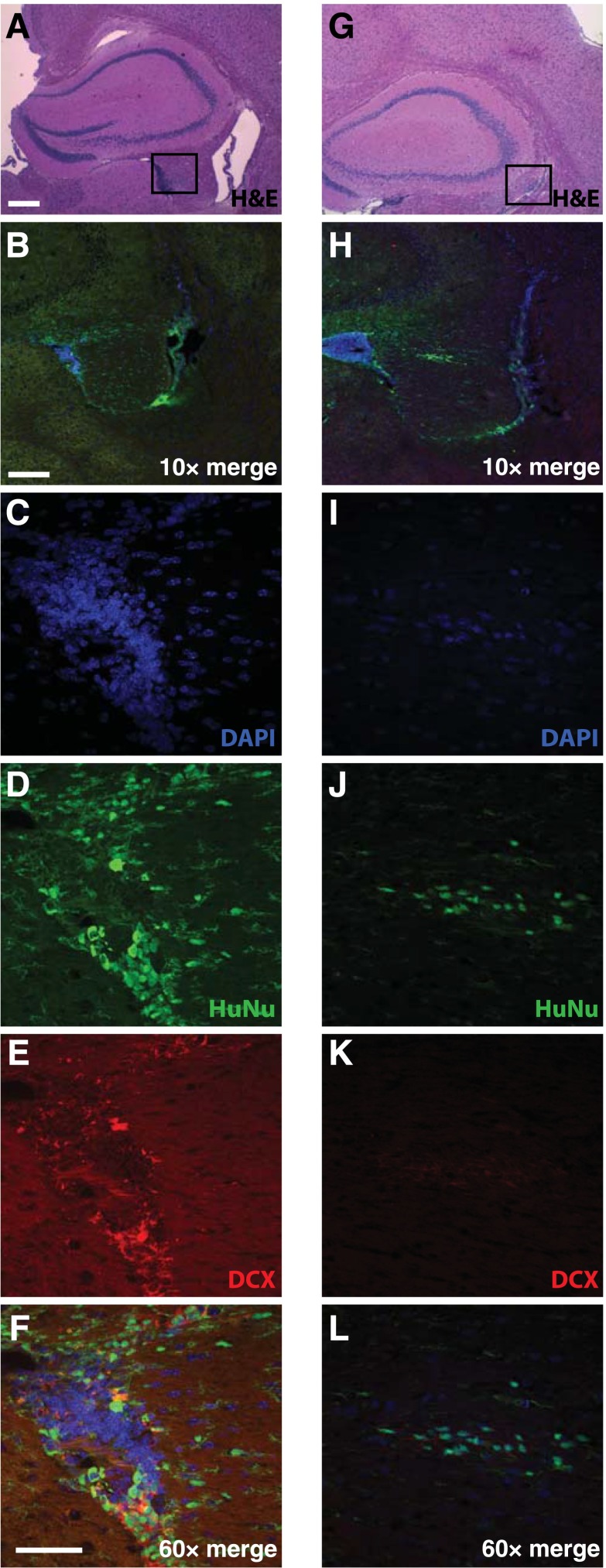
Alzheimer's disease (AD) is the most prevalent age-related neurodegenerative disorder and a leading cause of dementia. Current treatment fails to modify underlying disease pathologies and very little progress has been made to develop effective drug treatments. Cellular therapies impact disease by multiple mechanisms, providing increased efficacy compared with traditional single-target approaches. In amyotrophic lateral sclerosis, we have shown that transplanted spinal neural stem cells (NSCs) integrate into the spinal cord, form synapses with the host, improve inflammation, and reduce disease-associated pathologies. Our current goal is to develop a similar "best in class" cellular therapy for AD. Here, we characterize a novel human cortex-derived NSC line modified to express insulin-like growth factor-I (IGF-I), HK532-IGF-I. Because IGF-I promotes neurogenesis and synaptogenesis in vivo, this enhanced NSC line offers additional environmental enrichment, enhanced neuroprotection, and a multifaceted approach to treating complex AD pathologies. We show that autocrine IGF-I production does not impact the cell secretome or normal cellular functions, including proliferation, migration, or maintenance of progenitor status. However, HK532-IGF-I cells preferentially differentiate into gamma-aminobutyric acid-ergic neurons, a subtype dysregulated in AD; produce increased vascular endothelial growth factor levels; and display an increased neuroprotective capacity in vitro. We also demonstrate that HK532-IGF-I cells survive peri-hippocampal transplantation in a murine AD model and exhibit long-term persistence in targeted brain areas. In conclusion, we believe that harnessing the benefits of cellular and IGF-I therapies together will provide the optimal therapeutic benefit to patients, and our findings support further preclinical development of HK532-IGF-I cells into a disease-modifying intervention for AD.
Keywords: Alzheimer’s disease; Cellular therapy; Insulin-like growth factor-I; Neural stem cell; Neurodegeneration; Stem cell transplantation.
©AlphaMed Press.
Figures
Similar articles
-
Human neural stem cells improve cognition and promote synaptic growth in two complementary transgenic models of Alzheimer's disease and neuronal loss.Hippocampus. 2015 Jul;25(7):813-26. doi: 10.1002/hipo.22405. Epub 2015 Jan 8. Hippocampus. 2015. PMID: 25530343 Free PMC article.
-
Designer Self-Assemble Peptides Maximize the Therapeutic Benefits of Neural Stem Cell Transplantation for Alzheimer's Disease via Enhancing Neuron Differentiation and Paracrine Action.Mol Neurobiol. 2016 Mar;53(2):1108-1123. doi: 10.1007/s12035-014-9069-y. Epub 2015 Jan 14. Mol Neurobiol. 2016. PMID: 25586060 Free PMC article.
-
Autocrine production of IGF-I increases stem cell-mediated neuroprotection.Stem Cells. 2015 May;33(5):1480-9. doi: 10.1002/stem.1933. Stem Cells. 2015. PMID: 25532472
-
Effects of neural stem cell transplantation in Alzheimer's disease models.J Biomed Sci. 2020 Jan 27;27(1):29. doi: 10.1186/s12929-020-0622-x. J Biomed Sci. 2020. PMID: 31987051 Free PMC article. Review.
-
Neural stem cell-based treatment for neurodegenerative diseases.Neuropathology. 2013 Oct;33(5):491-504. doi: 10.1111/neup.12020. Epub 2013 Feb 5. Neuropathology. 2013. PMID: 23384285 Review.
Cited by
-
Advances in stromal cell therapy for management of Alzheimer's disease.Front Pharmacol. 2022 Oct 4;13:955401. doi: 10.3389/fphar.2022.955401. eCollection 2022. Front Pharmacol. 2022. PMID: 36267273 Free PMC article. Review.
-
Neural stem cells promote neuroplasticity: a promising therapeutic strategy for the treatment of Alzheimer's disease.Neural Regen Res. 2024 Mar;19(3):619-628. doi: 10.4103/1673-5374.380874. Neural Regen Res. 2024. PMID: 37721293 Free PMC article. Review.
-
Recent Advances: Decoding Alzheimer's Disease With Stem Cells.Front Aging Neurosci. 2018 Mar 22;10:77. doi: 10.3389/fnagi.2018.00077. eCollection 2018. Front Aging Neurosci. 2018. PMID: 29623038 Free PMC article. Review.
-
Human neural stem cells restore spatial memory in a transgenic Alzheimer's disease mouse model by an immunomodulating mechanism.Front Aging Neurosci. 2023 Dec 14;15:1306004. doi: 10.3389/fnagi.2023.1306004. eCollection 2023. Front Aging Neurosci. 2023. PMID: 38155736 Free PMC article.
-
In vivo and ex vivo gene therapy for neurodegenerative diseases: a promise for disease modification.Naunyn Schmiedebergs Arch Pharmacol. 2024 Oct;397(10):7501-7530. doi: 10.1007/s00210-024-03141-4. Epub 2024 May 22. Naunyn Schmiedebergs Arch Pharmacol. 2024. PMID: 38775852 Review.
References
-
- Thies W, Bleiler L. 2013 Alzheimer’s disease facts and figures. Alzheimers Dement. 2013;9:208–245. - PubMed
-
- Katzman R. Alzheimer’s disease. N Engl J Med. 1986;314:964–973. - PubMed
-
- LaFerla FM, Tinkle BT, Bieberich CJ, et al. The Alzheimer’s A beta peptide induces neurodegeneration and apoptotic cell death in transgenic mice. Nat Genet. 1995;9:21–30. - PubMed
-
- Selkoe DJ. Alzheimer’s disease is a synaptic failure. Science. 2002;298:789–791. - PubMed
Publication types
MeSH terms
Substances
Grants and funding
LinkOut - more resources
Full Text Sources
Other Literature Sources
Medical
