Case Reports
. 2016 Feb 25;127(8):997-1006.
doi: 10.1182/blood-2015-09-671636. Epub 2016 Jan 7.

Mutations in AP3D1 associated with immunodeficiency and seizures define a new type of Hermansky-Pudlak syndrome

Affiliations
Case Reports

Mutations in AP3D1 associated with immunodeficiency and seizures define a new type of Hermansky-Pudlak syndrome

Sandra Ammann et al. Blood. .

Abstract

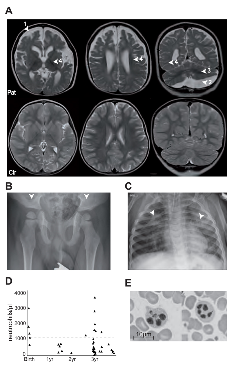
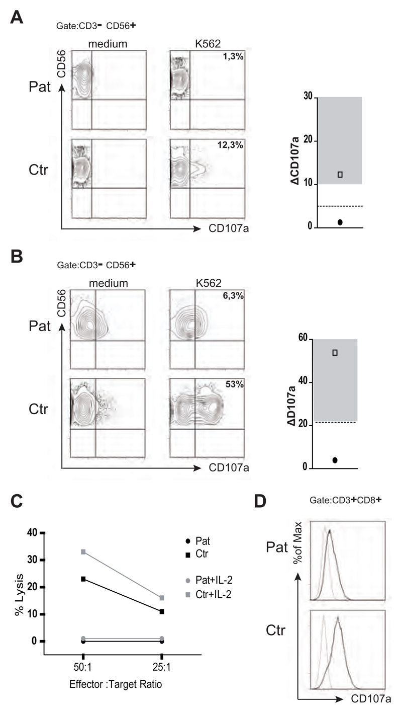
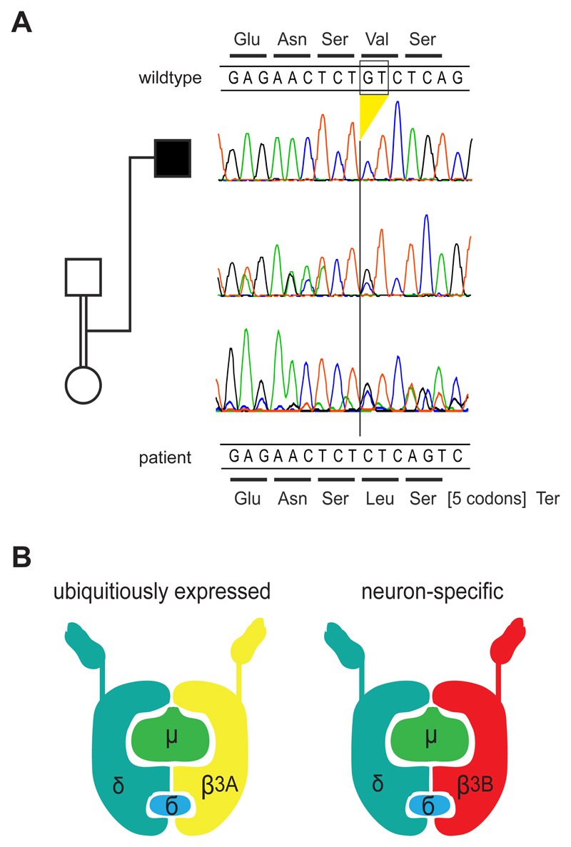
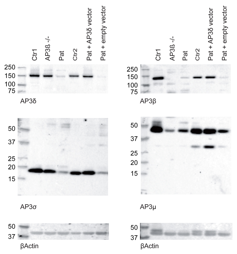
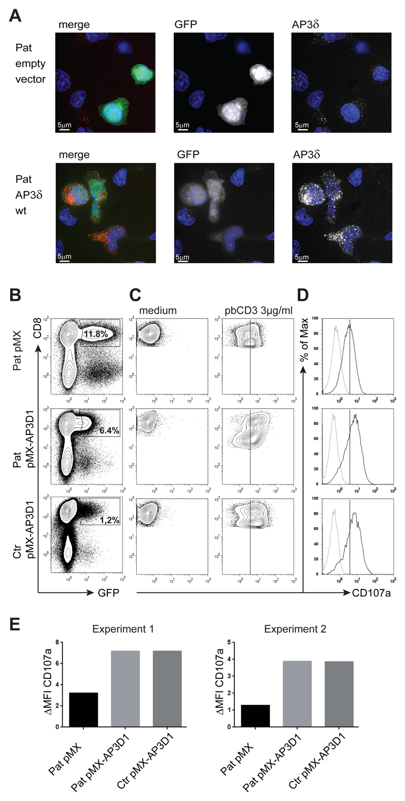
Genetic disorders affecting biogenesis and transport of lysosome-related organelles are heterogeneous diseases frequently associated with albinism. We studied a patient with albinism, neutropenia, immunodeficiency, neurodevelopmental delay, generalized seizures, and impaired hearing but with no mutation in genes so far associated with albinism and immunodeficiency. Whole exome sequencing identified a homozygous mutation in AP3D1 that leads to destabilization of the adaptor protein 3 (AP3) complex. AP3 complex formation and the degranulation defect in patient T cells were restored by retroviral reconstitution. A previously described hypopigmented mouse mutant with an Ap3d1 null mutation (mocha strain) shares the neurologic phenotype with our patient and shows a platelet storage pool deficiency characteristic of Hermansky-Pudlak syndrome (HPS) that was not studied in our patient because of a lack of bleeding. HPS2 caused by mutations in AP3B1A leads to a highly overlapping phenotype without the neurologic symptoms. The AP3 complex exists in a ubiquitous and a neuronal form. AP3D1 codes for the AP3δ subunit of the complex, which is essential for both forms. In contrast, the AP3β3A subunit, affected in HPS2 patients, is substituted by AP3β3B in the neuron-specific heterotetramer. AP3δ deficiency thus causes a severe neurologic disorder with immunodeficiency and albinism that we propose to classify as HPS10.


Conflict of interest statement

Disclosure of Conflicts of Interest

The authors declare they have no potential conflict of interest.

Figures

Figure 1
Figure 1. Clinical phenotype of the patient
(A) MRI scan of the brain at 3.5 years (upper panel), showing atrophy of the telencephalon (1), enlarged external and internal cerebrospinal fluid spaces (4), arachnoidal cyst (2) in the posterior fossa and insufficient myelination (3) indicated by arrow heads. The lower panel shows an MRI scan of a healthy 3.5 year old child. (B) X-ray of the pelvis showing flat, dyplastic acetabulae. (C) X-rays of the chest showing chronic interstitial pneumonia. (D) Neutrophil counts over time. Dashed line at 1000 neutrophiles/μl represent limit for neutropenia. (E) Bone marrow smear showing hypersegmented neutrophils.
Figure 2
Figure 2. Impaired degranulation and cytotoxicity of patient NK cells
(A) Ex vivo degranulation of NK cells (CD3-CD56+) from the patient (Pat) and a healthy donor (Ctr) after incubation with medium (left panel) or with K562 cells (middle panel) as assessed by flow cytometric analysis of CD107a surface expression. The right panel shows the difference in CD107a expression between unstimulated and stimulated cells of the patient (closed symbol) and a day control (open symbol) relative to healthy controls (gray area). The dashed line represents the level below which NK cell degranulation has the best positive and negative predictive value for a mutation in a gene relevant for cytotoxicity in an unfiltered cohort of patients with HLH. (B) Degranulation of NK cells prestimulated with IL-2 and PHA for 48 hours. (C) Cytotoxicity of patient (circle) and control (squares) PBMC on K562 target cells without (black symbols) or after overnight pre-incubation with IL-2 (grey symbols). (D) T cell degranulation. Cultured T cell of the patient and a healthy control were incubated in medium or stimulated with 3μg/ml of plate-bound anti-CD3. The increase in CD107a expression upon stimulation is shown as an overlay of histograms of unstimulated (dotted line) and stimulated (solid line) cells.
Figure 3
Figure 3. Genetic analysis and model of the AP-3 complex
(A) Electropherograms of the section of exon 32 harboring the homozygous deletion in the patient. The deletion leads to a frameshift at codon 1189 and a termination codon after seven residues. The encoded wild-type peptide sequence is shown at the top, the codons of the mutant sequence are shown below the DNA sequences. The parents are heterozygous for the deletion as expected. For technical reasons the reverse strand was sequenced in the DNA sample of the father but the complementary sequence is depicted here, i.e. it is shown in the same direction as the other samples. (B) Model of the AP-3 protein complex in the ubiquitous and the neuronal form (adapted from 31). AP-3δ is an essential part of both complexes, while AP-3β3A, mutated in HPS-2, is substituted by AP-3β3B in neuronal cells.
Figure 4
Figure 4. Reduced expression of AP-3δ affects the stability of the AP-3 complex
Western blot analysis of lysates from Macs-purified CD8 T cell of two healthy controls, an AP-3β3A deficient patient and the index patient (lane 1-4). Two separate blots are using rabbit antibodies against AP-3δ and AP-3σ (left) and AP-3β and AP-3μ (right). β-actin was used as a loading control. AP-3δ protein was analysed using two different antibodies, in three independent experiments each, each giving the same results. Lanes 5 and 6 show blots from patient PHA/IL-2 blasts retrovirally transduced with an AP3D1 expressing or empty vector analysed in the same experiments. These cells were FACS sorted for GFP positive cells to enrich for successfully transduced cells.
Figure 5
Figure 5. Genetic reconstitution with wild-type AP3D1 showing expression and function of the AP-3 complex
(A) Immunofluorescence analysis of patient PHA/IL-2 blasts reconstituted with either empty vector or wild-type AP3D1 using mouse anti-AP-3δ (SA4). Cells are shown in one 3D stack picture of 28 slices. Cells successfully transduced with the vector are GFP+ (white in the single stains, green in the merged picture), the nucleus is stained with Hoechst (blue) and AP-3δ is stained with SA4 provided by A. Peden (white in the single stains, red in the merged picture). The variable GFP intensity of reconstituted cells is explained by the use of an IRES-GFP construct where AP3D1 is placed before and GFP is placed after the IRES sequence. (B) Transduced T cells were rested in medium free of IL-2 for 24 hours and then incubated in medium or stimulated with 3μg/ml pb CD3 for 3 hours. Transduction efficiencies varied between 1 and 12%. (C) CD107a expression after incubation of transduced cells with medium or plate-bound anti-CD3. Plots are gated on successfully transduced GFP+ CD8+ cells. The solid line serves indicates peak CD107a expression in patient cells transduced with the empty vector. (D) Overlay of CD107a expression of transduced T cells kept in medium (dotted line) and stimulated stimulated with anti-CD3 (solid line) (E) The difference in mean fluorescence intensity (delta MFI) of CD107a expression between cells incubated in medium versus stimulated with pb anti-CD3 is shown for patient (pat) and ctonrol (ctr) GFP+CD8+ T cells transduced with the indicated vector. Results are shown from two independent experiments.

Comment in

References

    1. Dotta L, Parolini S, Prandini A, et al. Clinical, laboratory and molecular signs of immunodeficiency in patients with partial oculo-cutaneous albinism. Orphanet J Rare Dis. 2013;8:168. - PMC - PubMed
    1. Jessen B, Bode SF, Ammann S, et al. The risk of hemophagocytic lymphohistiocytosis in Hermansky-Pudlak syndrome type 2. Blood. 2013;121(15):2943–2951. - PMC - PubMed
    1. Griscelli C, Durandy A, Guy-Grand D, Daguillard F, Herzog C, Prunieras M. A syndrome associating partial albinism and immunodeficiency. Am J Med. 1978;65(4):691–702. - PubMed
    1. Blume RS, Wolff SM. The Chediak-Higashi syndrome: studies in four patients and a review of the literature. Medicine (Baltimore) 1972;51(4):247–280. - PubMed
    1. Dell'Angelica EC, Shotelersuk V, Aguilar RC, Gahl WA, Bonifacino JS. Altered trafficking of lysosomal proteins in Hermansky-Pudlak syndrome due to mutations in the beta 3A subunit of the AP-3 adaptor. Mol Cell. 1999;3(1):11–21. - PubMed

Publication types

MeSH terms

Substances