. 2015 Dec 15:4:e11190.
doi: 10.7554/eLife.11190.

Hippocampus ghrelin signaling mediates appetite through lateral hypothalamic orexin pathways

Affiliations

Hippocampus ghrelin signaling mediates appetite through lateral hypothalamic orexin pathways

Ted M Hsu et al. Elife. .

Abstract

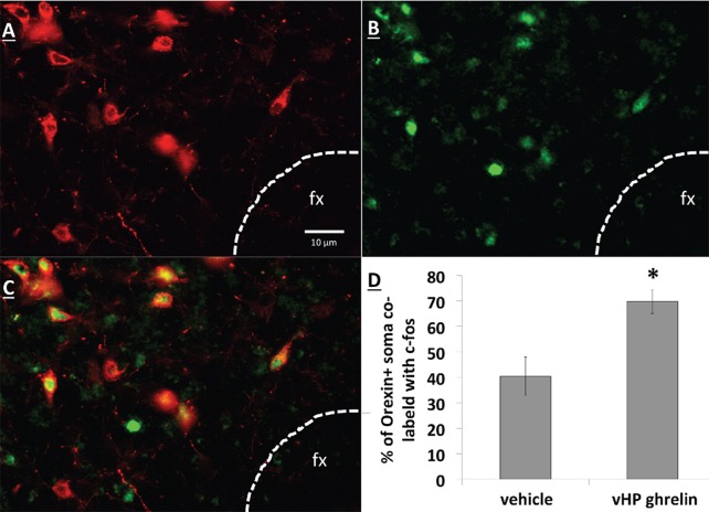
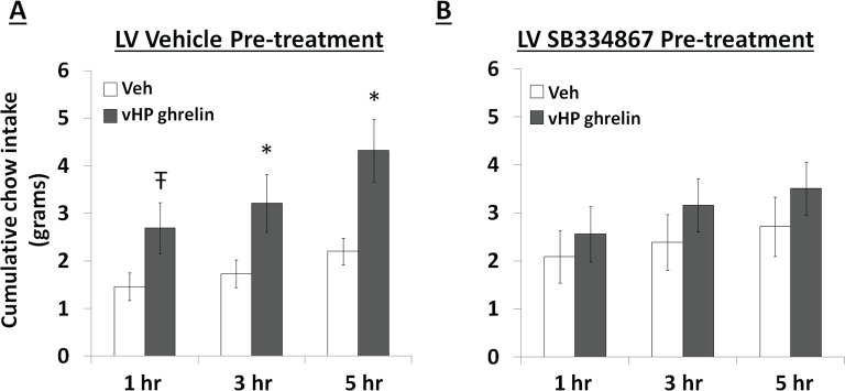
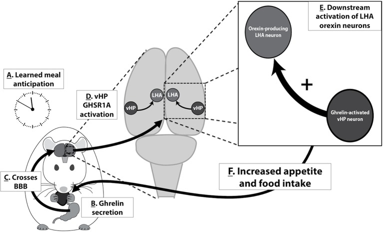
Feeding behavior rarely occurs in direct response to metabolic deficit, yet the overwhelming majority of research on the biology of food intake control has focused on basic metabolic and homeostatic neurobiological substrates. Most animals, including humans, have habitual feeding patterns in which meals are consumed based on learned and/or environmental factors. Here we illuminate a novel neural system regulating higher-order aspects of feeding through which the gut-derived hormone ghrelin communicates with ventral hippocampus (vHP) neurons to stimulate meal-entrained conditioned appetite. Additional results show that the lateral hypothalamus (LHA) is a critical downstream substrate for vHP ghrelin-mediated hyperphagia and that vHP ghrelin activated neurons communicate directly with neurons in the LHA that express the neuropeptide, orexin. Furthermore, activation of downstream orexin-1 receptors is required for vHP ghrelin-mediated hyperphagia. These findings reveal novel neurobiological circuitry regulating appetite through which ghrelin signaling in hippocampal neurons engages LHA orexin signaling.

Keywords: GHSR; appetite; feeding; learning; neuroscience; obesity; rat; ventral hippocampus.


Conflict of interest statement

The authors declare that no competing interests exist.

Figures

Figure 1.
Figure 1.. Representative vHP injection site is shown as localization of pontamine sky blue ink following a 100 nl injection.
CA1sp, CA3sp: CA1 and CA3 pyramidal cells; alv = alveus. DOI: http://dx.doi.org/10.7554/eLife.11190.003
Figure 2.
Figure 2.. Effects of vHP GHSR blockade on conditioned food intake.
(A) Daily 4 hr chow intake during meal entrainment acquisition, where animals were limited to 4 hr food intake/day for 8 days. (B) 4 hr chow intake in meal-entrained animals following bilateral vHP administration of JMV2959. (C) 4 hr chow intake in non-meal entrained, 20 hr food-restricted rats following bilateral vHP administration of JMV2959. (D) 4 hr Western diet intake in non-meal entrained, non-food deprived rats following bilateral vHP administration of JMV2959. Data are mean ± SEM; *p<0.05. DOI: http://dx.doi.org/10.7554/eLife.11190.004
Figure 2—figure supplement 1.
Figure 2—figure supplement 1.. Effects of LHA GHSR blockade on conditioned food intake.
(A) Daily 4 hr chow intake during meal entrainment acquisition. (B) 4 hr chow intake in meal-entrained animals following bilateral LHA administration of JMV2959. (C) 4 hr chow intake in non-meal entrained, 20 hr food-restricted rats following bilateral LHA administration of JMV2959. Data are mean ± SEM. DOI: http://dx.doi.org/10.7554/eLife.11190.005
Figure 3.
Figure 3.. GHSR-expressing vHP CA1 neurons provide input to dpLHA neurons.
(A) A representative dorsal perifornical LHA injection site of CTB-AF594 (red). fx:fornix; V3h: Third ventricle (B) Darkfield cytoarchitecture of the vHP, where insets C-E represents field CA1 pyramidal layer (CA1sp) insets from the square (white dashed line). (C) vHP retrogradely-labeled CTB immunoreactive perikarya (red). (D) GHSR protein expression (green). (E) CTB and GHSR co-labeled soma. DOI: http://dx.doi.org/10.7554/eLife.11190.006
Figure 4.
Figure 4.. Representative unilateral dpLHA lesions.
(A) NeuN immunohistochemistry contralateral to a NMDA dorsal perifornical (dp) LHA lesion (intact side). (B) NeuN immunohistochemistry for a representative unilateral NMDA dpLHA lesion targeting the dorsal perifornical LHA. DOI: http://dx.doi.org/10.7554/eLife.11190.007
Figure 5.
Figure 5.. Effects of vHP-dpLHA unilateral disconnection on vHP ghrelin-mediated hyperphagia.
Cumulative chow intake following unilateral vHP injections of ghrelin (300 pmol) that was: (A) Ipsilateral to a unilateral dpLHA lesion, (B) ipsilateral to a dpLHA sham lesion. (C) contralateral to a dpLHA lesion, and (D) contralateral to a dpLHA sham lesion. Data are mean ± SEM; *p<0.05; Ŧ p<0.07. DOI: http://dx.doi.org/10.7554/eLife.11190.008
Figure 6.
Figure 6.. vHP CA1 neurons provide input to dpLHA ORX-expressing neurons.
(A) A representative PHAL (blue) injection site centered in ventral hippocampus field CA1. CA1sp, CA3sp: CA1 and CA3 pyramidal cells. (B) Darkfield microscopy, where neuroanatomical analyses were performed in the dpLHA (fx: fornix; V3h: Third ventricle; location shown in C). Inset (C) confocal imaging reveals some orexin neurons (red) in the dpLHA that are in close apposition to vHP field CA1-originating axons, labeled with PHAL (blue). (D) Triple-label imaging reveals co-labeling of PHAL and synaptophysin (green) in axons in apposition to dpLHA orexin-expressing soma (indicated by white arrows). DOI: http://dx.doi.org/10.7554/eLife.11190.009
Figure 6—figure supplement 1.
Figure 6—figure supplement 1.. CA1 vHP neurons project ipsilaterally to the dpLHA.
(A–C) PHAL-immunoreactive axons originating from the vHP (field CA1) within the dpLHA, revealing an exclusively ipsilateral projection from the vHP to the dpLHA (v3h = 3rd ventricle; fx = fornix). DOI: http://dx.doi.org/10.7554/eLife.11190.010
Figure 7.
Figure 7.. vHP GHSR activation increases Fos expression in dpLHA orexin-expressing neurons.
(A) Orexin-expressing neurons (red) in the dpLHA; fx: fornix.  (B) Fos expression (green) in the dpLHA induced by ipsilateral vHP ghrelin (300 pmol) administration. (C) Co-labeling of dpLHA orexin expression and Fos expression. (D) Quantification of orexin positive soma co-labeled with Fos, where co-labeling was significantly higher compared to vHP vehicle injections. Data are mean ± SEM; *p<0.05. DOI: http://dx.doi.org/10.7554/eLife.11190.011
Figure 8.
Figure 8.. Effects of Orexin-1 receptor blockade on ghrelin-mediated hyperphagia.
Cumulative chow intake following: (A) pre-treatment with lateral ventricle (LV) vehicle followed by vHP ghrelin (300 pmol, unilateral) administration or (B) LV Ox1R antagonist, SB334867 (30 nmol), followed by vHP ghrelin administration. Data are mean ± SEM; *p<0.05; Ŧ p<0.07. DOI: http://dx.doi.org/10.7554/eLife.11190.012
Figure 9.
Figure 9.. A model for vHP ghrelin-mediated conditioned feeding behavior.
(A) Animals learn to anticipate a meal based on external or internal food cues. (B,C) Ghrelin secreted from the stomach in anticipation of feeding crosses the blood brain barrier and enters the central nervous system. (D) Ghrelin acts on GHSR in vHP neurons, which engages (E) Downstream activation of dpLHA orexin neurons and CNS Ox1R signaling. (F) Activation of this neural pathway increases conditioned appetite and feeding behavior. DOI: http://dx.doi.org/10.7554/eLife.11190.013

Similar articles

Cited by

References

    1. Alvarez-Crespo M, Skibicka KP, Farkas I, Molnár CS, Egecioglu E, Hrabovszky E, Liposits Z, Dickson SL. The amygdala as a neurobiological target for ghrelin in rats: neuroanatomical, electrophysiological and behavioral evidence. PloS One. 2012;7:e11190. doi: 10.1371/journal.pone.0046321. - DOI - PMC - PubMed
    1. Anand BK, Brobeck JR. Hypothalamic control of food intake in rats and cats. The Yale Journal of Biology and Medicine. 1951;24:123–140. - PMC - PubMed
    1. Benoit SC, Davis JF, Davidson TL. Learned and cognitive controls of food intake. Brain Research. 2010;1350:71–76. doi: 10.1016/j.brainres.2010.06.009. - DOI - PMC - PubMed
    1. Berthoud HR, Münzberg H. The lateral hypothalamus as integrator of metabolic and environmental needs: from electrical self-stimulation to opto-genetics. Physiology & Behavior. 2011;104:29–39. doi: 10.1016/j.physbeh.2011.04.051. - DOI - PMC - PubMed
    1. Blum ID, Patterson Z, Khazall R, Lamont EW, Sleeman MW, Horvath TL, Abizaid A. Reduced anticipatory locomotor responses to scheduled meals in ghrelin receptor deficient mice. Neuroscience. 2009;164:351–359. doi: 10.1016/j.neuroscience.2009.08.009. - DOI - PMC - PubMed

Publication types