

Expression of Human NSAID Activated Gene 1 in Mice Leads to Altered Mammary Gland Differentiation and Impaired Lactation
- PMID: 26745373
- PMCID: PMC4706436
- DOI: 10.1371/journal.pone.0146518
Expression of Human NSAID Activated Gene 1 in Mice Leads to Altered Mammary Gland Differentiation and Impaired Lactation
Erratum in
-
Correction: Expression of Human NSAID Activated Gene 1 in Mice Leads to Altered Mammary Gland Differentiation and Impaired Lactation.PLoS One. 2016 Mar 10;11(3):e0151504. doi: 10.1371/journal.pone.0151504. eCollection 2016. PLoS One. 2016. PMID: 26963910 Free PMC article. No abstract available.
Abstract
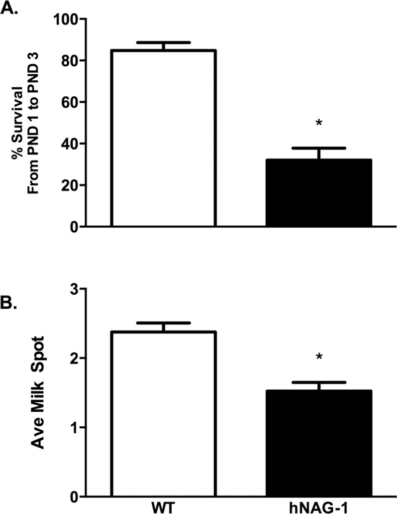
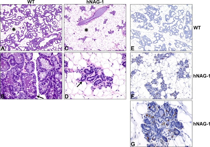
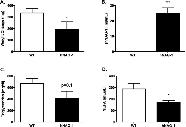
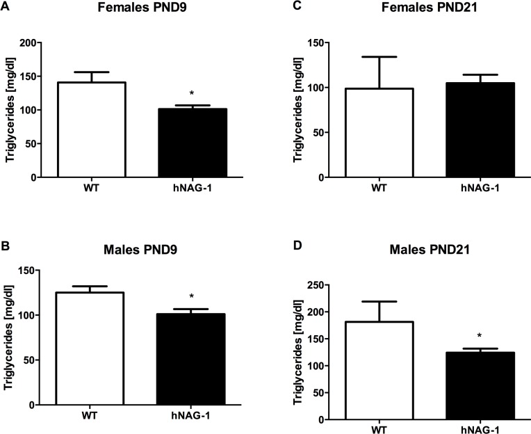
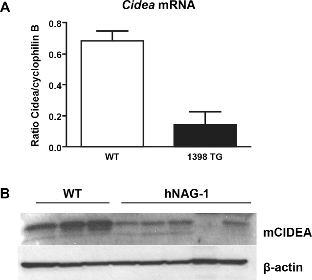
Transgenic mice expressing human non-steroidal anti-inflammatory drug activated gene 1 (NAG-1) have less adipose tissue, improved insulin sensitivity, lower insulin levels and are resistant to dietary induced obesity. The hNAG-1 expressing mice are more metabolically active with a higher energy expenditure. This study investigates female reproduction in the hNAG-1 transgenic mice and finds the female mice are fertile but have reduced pup survival after birth. Examination of the mammary glands in these mice suggests that hNAG-1 expressing mice have altered mammary epithelial development during pregnancy, including reduced occupancy of the fat pad and increased apoptosis via TUNEL positive cells on lactation day 2. Pups nursing from hNAG-1 expressing dams have reduced milk spots compared to pups nursing from WT dams. When CD-1 pups were cross-fostered with hNAG-1 or WT dams; reduced milk volume was observed in pups nursing from hNAG-1 dams compared to pups nursing from WT dams in a lactation challenge study. Milk was isolated from WT and hNAG-1 dams, and the milk was found to have secreted NAG-1 protein (approximately 25 ng/mL) from hNAG-1 dams. The WT dams had no detectable hNAG-1 in the milk. A decrease in non-esterified free fatty acids in the milk of hNAG-1 dams was observed. Altered milk composition suggests that the pups were receiving inadequate nutrients during perinatal development. To examine this hypothesis serum was isolated from pups and clinical chemistry points were measured. Male and female pups nursing from hNAG-1 dams had reduced serum triglyceride concentrations. Microarray analysis revealed that genes involved in lipid metabolism are differentially expressed in hNAG-1 mammary glands. Furthermore, the expression of Cidea/CIDEA that has been shown to regulate milk lipid secretion in the mammary gland was reduced in hNAG-1 mammary glands. This study suggests that expression of hNAG-1 in mice leads to impaired lactation and reduces pup survival due to altered milk quality and quantity.
Conflict of interest statement
Figures
Similar articles
-
Reduced milk triglycerides in mice lacking phosphoenolpyruvate carboxykinase in mammary gland adipocytes and white adipose tissue contribute to the development of insulin resistance in pups.J Nutr. 2009 Dec;139(12):2257-65. doi: 10.3945/jn.109.113092. Epub 2009 Oct 7. J Nutr. 2009. PMID: 19812223 Free PMC article.
-
NTP technical report on the toxicity studies of Dibutyl Phthalate (CAS No. 84-74-2) Administered in Feed to F344/N Rats and B6C3F1 Mice.Toxic Rep Ser. 1995 Apr;30:1-G5. Toxic Rep Ser. 1995. PMID: 12209194
-
NAG-1/GDF-15 prevents obesity by increasing thermogenesis, lipolysis and oxidative metabolism.Int J Obes (Lond). 2014 Dec;38(12):1555-64. doi: 10.1038/ijo.2014.27. Epub 2014 Feb 17. Int J Obes (Lond). 2014. PMID: 24531647 Free PMC article.
-
NIEHS Report on the Toxicity Studies of Nicotine Bitartrate Dihydrate (CASRN 6019-06-3) Administered in Drinking Water to Sprague Dawley Rats and Swiss Mice: NIEHS Report 11 [Internet].Research Triangle Park (NC): National Institute of Environmental Health Sciences; 2024 Oct. Research Triangle Park (NC): National Institute of Environmental Health Sciences; 2024 Oct. PMID: 39441944 Free Books & Documents. Review.
-
Review: Mammary gland development in swine: embryo to early lactation.Animal. 2019 Jul;13(S1):s11-s19. doi: 10.1017/S1751731119000521. Animal. 2019. PMID: 31280748 Review.
Cited by
-
A regulatory T cell Notch4-GDF15 axis licenses tissue inflammation in asthma.Nat Immunol. 2020 Nov;21(11):1359-1370. doi: 10.1038/s41590-020-0777-3. Epub 2020 Sep 14. Nat Immunol. 2020. PMID: 32929274 Free PMC article.
-
GDF15 Knockout Does Not Substantially Impact Perinatal Body Weight or Neonatal Outcomes in Mice.Endocrinology. 2024 Oct 30;165(12):bqae143. doi: 10.1210/endocr/bqae143. Endocrinology. 2024. PMID: 39445824
-
GDF15 knockout does not substantially impact perinatal body weight or neonatal outcomes in mice.bioRxiv [Preprint]. 2024 May 3:2024.04.30.591359. doi: 10.1101/2024.04.30.591359. bioRxiv. 2024. Update in: Endocrinology. 2024 Oct 30;165(12):bqae143. doi: 10.1210/endocr/bqae143. PMID: 38746399 Free PMC article. Updated. Preprint.
-
Correction: Expression of Human NSAID Activated Gene 1 in Mice Leads to Altered Mammary Gland Differentiation and Impaired Lactation.PLoS One. 2016 Mar 10;11(3):e0151504. doi: 10.1371/journal.pone.0151504. eCollection 2016. PLoS One. 2016. PMID: 26963910 Free PMC article. No abstract available.
-
Nutritional Programming of Bone Structure in Male Offspring by Maternal Consumption of Citrus Flavanones.Calcif Tissue Int. 2018 Jun;102(6):671-682. doi: 10.1007/s00223-017-0366-0. Epub 2017 Nov 18. Calcif Tissue Int. 2018. PMID: 29151126 Free PMC article.
References
-
- Daniel CW, Silberstein GB, Strickland P. Direct action of 17 beta-estradiol on mouse mammary ducts analyzed by sustained release implants and steroid autoradiography. Cancer research. 1987;47(22):6052–7. Epub 1987/11/15. . - PubMed
-
- Hewitt SC, Bocchinfuso WP, Zhai J, Harrell C, Koonce L, Clark J, et al. Lack of ductal development in the absence of functional estrogen receptor alpha delays mammary tumor formation induced by transgenic expression of ErbB2/neu. Cancer research. 2002;62(10):2798–805. Epub 2002/05/23. . - PubMed
-
- Hewitt SC, Kissling GE, Fieselman KE, Jayes FL, Gerrish KE, Korach KS. Biological and biochemical consequences of global deletion of exon 3 from the ER alpha gene. FASEB journal: official publication of the Federation of American Societies for Experimental Biology. 2010;24(12):4660–7. Epub 2010/07/30. 10.1096/fj.10-163428 ; PubMed Central PMCID: PMCPmc2992373. - DOI - PMC - PubMed
-
- Humphreys RC, Lydon JP, O'Malley BW, Rosen JM. Use of PRKO mice to study the role of progesterone in mammary gland development. Journal of mammary gland biology and neoplasia. 1997;2(4):343–54. Epub 2000/08/10. . - PubMed
Publication types
MeSH terms
Substances
Grants and funding
LinkOut - more resources
Full Text Sources
Other Literature Sources
Molecular Biology Databases
