. 2016 Jan 8;10(1):e0004298.
doi: 10.1371/journal.pntd.0004298. eCollection 2016 Jan.

In Vitro Mode of Action and Anti-thrombotic Activity of Boophilin, a Multifunctional Kunitz Protease Inhibitor from the Midgut of a Tick Vector of Babesiosis, Rhipicephalus microplus

Affiliations

In Vitro Mode of Action and Anti-thrombotic Activity of Boophilin, a Multifunctional Kunitz Protease Inhibitor from the Midgut of a Tick Vector of Babesiosis, Rhipicephalus microplus

Teresa C Assumpção et al. PLoS Negl Trop Dis. .

Abstract

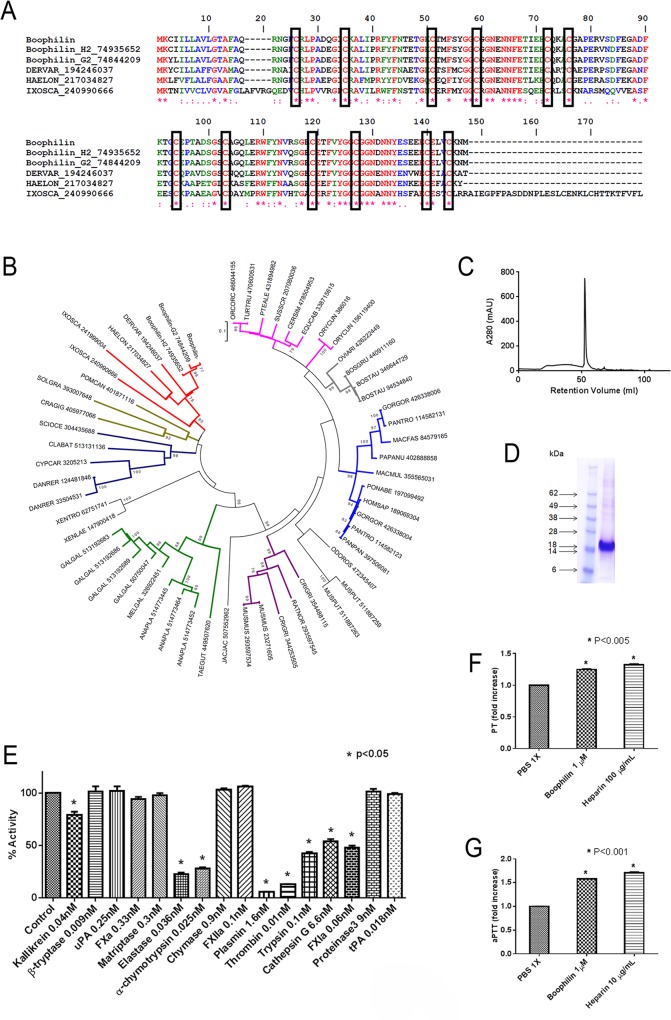
Background: Hematophagous mosquitos and ticks avoid host hemostatic system through expression of enzyme inhibitors targeting proteolytic reactions of the coagulation and complement cascades. While most inhibitors characterized to date were found in the salivary glands, relatively few others have been identified in the midgut. Among those, Boophilin is a 2-Kunitz multifunctional inhibitor targeting thrombin, elastase, and kallikrein. However, the kinetics of Boophilin interaction with these enzymes, how it modulates platelet function, and whether it inhibits thrombosis in vivo have not been determined.

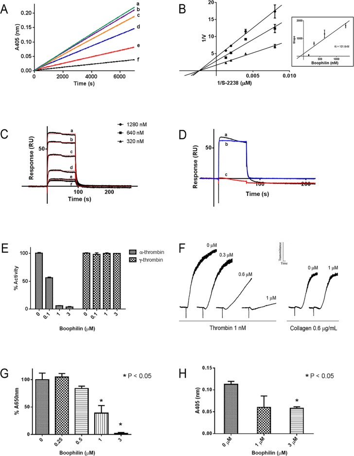
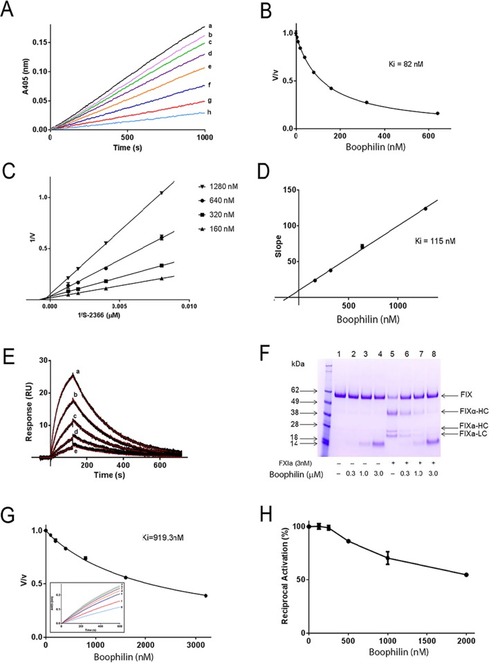
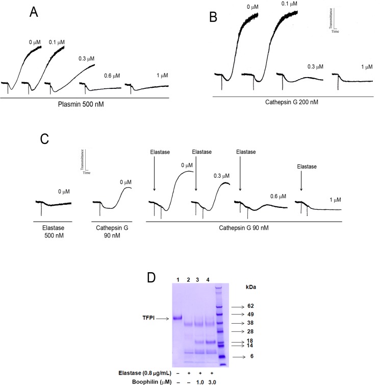
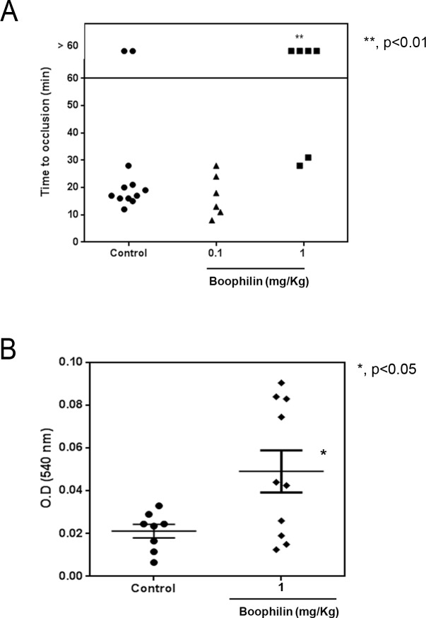
Methodology/principal findings: Boophilin was expressed in HEK293 cells and purified to homogeneity. Using amidolytic assays and surface plasmon resonance experiments, we have demonstrated that Boophilin behaves as a classical, non-competitive inhibitor of thrombin with respect to small chromogenic substrates by a mechanism dependent on both exosite-1 and catalytic site. Inhibition is accompanied by blockade of platelet aggregation, fibrin formation, and clot-bound thrombin in vitro. Notably, we also identified Boophilin as a non-competitive inhibitor of FXIa, preventing FIX activation. In addition, Boophilin inhibits kallikrein activity and the reciprocal activation, indicating that it targets the contact pathway. Furthermore, Boophilin abrogates cathepsin G- and plasmin-induced platelet aggregation and partially affects elastase-mediated cleavage of Tissue Factor Pathway Inhibitor (TFPI). Finally, Boophilin inhibits carotid artery occlusion in vivo triggered by FeCl3, and promotes bleeding according to the mice tail transection method.

Conclusion/significance: Through inhibition of several enzymes involved in proteolytic cascades and cell activation, Boophilin plays a major role in keeping the midgut microenvironment at low hemostatic and inflammatory tonus. This response allows ticks to successfully digest a blood meal which is critical for metabolism and egg development. Boophilin is the first tick midgut FXIa anticoagulant also found to inhibit thrombosis.


Conflict of interest statement

The authors have declared that no competing interests exist.

Figures

Fig 1
Fig 1. Characterization of Boophilin.
(A) Clustal alignment of Boophilin [3,21] with other proteins of the Kunitz family. The boxes indicate the 12 conserved cysteines. The symbols below the alignment indicate: (*) identical sites; (:) conserved sites; (.) less conserved sites. (B) Phylogram of Boophilin and other Kunitz proteins, obtained by the Neighbor-joining algorithm, using pairwise deletion and poisson model. The sequences from the nonreduntant (NR) protein database of the National Center for Biotechnology Information (NCBI) are represented by the first 3 letters of their genus name, followed by the first three letters of the species name, followed by their gi| accession number. The numbers in the phylogram nodes indicate percent bootstrap support for the phylogeny after 10,000 iterations. The bar indicates 10% amino acid divergence in the sequences. (C) Reverse-phase chromatography of Boophilin expressed in HEK293 cells. (D) Boophilin was loaded in a NuPAGE gel under reducing conditions. Gel was stained with Coomassie Blue. On the left, molecular mass markers are indicated. (E) Protease screening. Screening for inhibition of several enzymes by Boophilin (100 nM): catalytic activity was estimated by fluorogenic substrate hydrolysis. (F) PT. Boophilin at 1 μM was added to the plasma, followed by addition of PT reagent as described in Methods. PT time, control human plasma: 17.5±0.25 seconds. (G) aPTT. Boophilin at 1 μM was added to the plasma, followed by addition of aPTT reagent as described in Methods. aPTT, control human plasma: 44.9±0.85 seconds. Clotting was estimated using a coagulometer and heparin was used as a control.
Fig 2
Fig 2. Boophilin inhibits thrombin.
(A) Typical progress curves for thrombin-mediated hydrolysis of S2238 in the absence and presence of Boophilin at the indicated concentrations (a, 0 nM; b, 20 nM; c; 40 nM; d, 80 nM; e, 160 nM; f, 320 nM). Reactions started with addition of S2238 (250 μM) to a mixture containing Boophilin incubated with thrombin (0.2 nM) for 30 minutes. Substrate hydrolysis was followed for 2 hours at 37°C and 405 nm. (B) Lineweaver-Burk plot of the inhibition of thrombin by Boophilin (320 nM, 640 nM, and 1280 nM) at different concentrations of substrate S2238. Inset: Re-plot of the slope versus Boophilin concentration. (C) SPR experiments. Thrombin at the indicated concentrations (a, 500 nM; b, 250 nM; c; 125 nM; d, 62.5 nM; e, 31.25 nM; f, 15.625 nM) was injected over immobilized Boophilin for 90 seconds. Dissociation of the Boophilin-thrombin complex was monitored for 180 seconds, and a global 1:1 binding model was used to calculate kinetics parameters. Representative sensorgrams are shown in black lines and fitting of the data points using the Langmuir equation is depicted in red lines. (D) Boophilin binds to α-thrombin (a) and PPACK-thrombin (b) but does not interact with γ-thrombin (c). All the analytes were tested at 1 μM and injected in a sensor chip containing immobilized Boophilin. (E) α-thrombin or γ-thrombin mediated hydrolysis of S2238 in the presence of Boophilin at the indicated concentrations. Reactions started with addition of S2238 (250 μM) to a mixture containing Boophilin incubated with α-thrombin (0.2 nM) or γ-thrombin (0.2 nM) for 30 minutes. Substrate hydrolysis was followed at 405 nm. (F) Platelet aggregation. Washed platelets were stimulated by thrombin (1 nM) in the absence or presence of Boophilin (0.3 μM, 0.6 μM, and 1.0 μM). Control: collagen-induced platelet aggregation. (G) Fibrinogen clotting activity. Thrombin (200 nM) was incubated with different concentrations of Boophilin for 10 minutes at 37°C. Fibrinogen (final concentration: 2 mg/mL) was added and absorbance at 600nm was recorded for 30 minutes, at 30 seconds intervals. (H) Clot-bound α-thrombin. Fibrin clots were incubated with 1 μM and 3 μM of Boophilin or PBS 1X. Chromogenic substrate (S2238) hydrolysis was estimated by end point reading at 405nm.
Fig 3
Fig 3. Boophilin inhibits FXIa and Kallikrein.
(A) Typical progress curves for FXIa-mediated S2366 hydrolysis in the absence and presence of Boophilin at the indicated concentrations (a, 0 nM; b, 10 nM; c, 20 nM; d, 40 nM; e, 80 nM; f, 160 nM; g, 320 nM; h, 640 nM). Reactions started with addition of S2366 (250 μM) to a mixture containing Boophilin incubated for 1 hour with FXIa (10 nM). Substrate hydrolysis was followed for 2 hours at 37°C and 405 nm. (B) The ratio of Vs/Vo in (A) was plotted against Boophilin concentration to calculate the apparent Ki. (C) Lineweaver-Burk plot of the inhibition of FXIa activity by Boophilin (160 nM, 320 nM, 640 nM, and 1280 nM) at different concentrations of substrate S2366. (D) Re-plot of the slope from Lineweaver-Burk plot versus Boophilin concentrations. (E) SPR experiments. Boophilin at different concentrations (a, 500 nM; b, 250 nM; c; 125 nM; d, 62.5 nM; e, 31.25 nM) was injected over immobilized FXIa for 120 seconds. Dissociation of the Boophilin-FXIa complex was monitored for 600 seconds, and a global 1:1 binding model was used to calculate kinetics parameters. Representative sensorgrams are shown in black lines, and fitting of the data points using the Langmuir equation is depicted in red lines. (F) Inhibition of FIX activation. Control (lanes 1, 2, 3, 4): recombinant FIX (BeneFIX, 1.0 μM) in the presence of PBS (lane 1) or indicated concentrations of Boophilin (lanes 2, 3, 4). Lanes 5, 6, 7 and 8: FXIa (3 nM) was added to FIX in the presence of PBS (lane 5) or Boophilin (lanes 6, 7, 8), and mixture was incubated at 37°C for 60 min. Reactions were stopped with reducing Laemmli buffer, and proteins were separated by 4% to 12% NuPAGE. The bands correspond to (from the top): uncleaved FIX (FIX), the heavy chain FIXα (FIXα-HC), the heavy chain of FIXa (FIXa-HC), and the light chain of FIXa (FIXa-LC). (G) The ratio of kallikrein activity (Vs/Vo) was plotted against Boophilin concentration. Inset: typical progress curves for kallikrein-mediated hydrolysis of S2302 in the absence and presence of Boophilin at the indicated concentrations (a, 0 nM; b, 100 nM; c; 200 nM; d, 400 nM; e, 800 nM; f, 1600 nM; g, 3200 nM). Reactions started with addition of S2302 (250 μM) to a mixture containing Boophilin incubated with kallikrein (2 nM) for 1 hour. Substrate hydrolysis was followed for 2 hours at 37°C and 405 nm. (H) Reciprocal activation. Factor XII (0.2 nM) was preincubated with Boophilin (0 nM, 125 nM, 250 nM, 500 nM, 1000 nM and 2000 nM) in 20 mM Tris, 0.15 M NaCl, 0.3% BSA, pH 7.4, for 10 minutes at room temperature. Reactions were started by addition of pre-kallikrein (10 nM) and DS 500 (0.2 μg/mL, final concentrations). Data points are the mean+/-SD (n = 3).
Fig 4
Fig 4. Boophilin inhibits plasmin, cathepsin G-induced platelet aggregation and elastase-mediated cleavage of TFPI.
(A) Washed platelets were stimulated by plasmin (500 nM) in the absence or presence of Boophilin (0.1 μM, 0.3 μM, 0.6 μM, and 1.0 μM). (B) Washed platelets were stimulated by Cathepsin G (200 nM) in the absence or presence of Boophilin (0.1 μM, 0.3 μM, and 1.0 μM). (C) Washed platelets were induced to aggregate by Elastase (500 nM) and Cathepsin G (90 nM) with the indicated concentrations of Boophilin. (D) TFPI Cleavage. Boophilin (0 μM, 1 μM, and 3 μM) was incubated with TFPI (1 μg), in the presence of elastase (final concentration: 0.8 μg/mL), for 2 hours at room temperature. Reactions were stopped by addition of Laemmli buffer and boiling with dithiothreitol for 5 minutes. Proteins were separated by 4% to 12% NuPAGE (MES buffer) and gel was stained with Coomassie Blue R-250. Control: TFPI only.
Fig 5
Fig 5. Boophilin inhibits thrombosis in vivo.
(A) Thrombosis in vivo. A paper filter imbibed with 5% FeCl3 was applied to the carotid artery, and blood flow was monitored with a perivascular flow probe for 60 minutes or until stable occlusion took place. Fifteen minutes before injury, Boophilin was injected into the caudal vein of the mice. Each symbol represents one individual. **, p < 0.01. (B) Bleeding time. Bleeding was caused by a tail transection after i.v. injection of Boophilin at 1 mg/Kg. Absorbance at 540 nm was used to estimate blood loss. *, p<0.05 (ANOVA, with Dunnett post-test).

Similar articles

Cited by

References

    1. Corral-Rodriguez MA, Macedo-Ribeiro S, Barbosa Pereira PJ, Fuentes-Prior P (2009) Tick-derived Kunitz-type inhibitors as antihemostatic factors. Insect Biochem Mol Biol 39: 579–595. 10.1016/j.ibmb.2009.07.003 - DOI - PubMed
    1. Koh CY, Kini RM (2009) Molecular diversity of anticoagulants from haematophagous animals. Thromb Haemost 102: 437–453. 10.1160/TH09-04-0221 - DOI - PubMed
    1. Louw E, van der Merwe NA, Neitz AW, Maritz-Olivier C (2012) Evolution of the tissue factor pathway inhibitor-like Kunitz domain-containing protein family in Rhipicephalus microplus. Int J Parasitol. - PubMed
    1. Francischetti IM, Sa-Nunes A, Mans BJ, Santos IM, Ribeiro JM (2009) The role of saliva in tick feeding. Front Biosci 14: 2051–2088. - PMC - PubMed
    1. Ribeiro JM, Francischetti IM (2003) Role of arthropod saliva in blood feeding: sialome and post-sialome perspectives. Annu Rev Entomol 48: 73–88. - PubMed

Publication types

MeSH terms

LinkOut - more resources