. 2017 Jan 2;11(1):13-23.
doi: 10.1080/19336918.2015.1134404. Epub 2016 Jan 8.

LPS-induced CXCR4-dependent migratory properties and a mesenchymal-like phenotype of colorectal cancer cells

Affiliations

LPS-induced CXCR4-dependent migratory properties and a mesenchymal-like phenotype of colorectal cancer cells

Wen-Ting Liu et al. Cell Adh Migr. .

Abstract

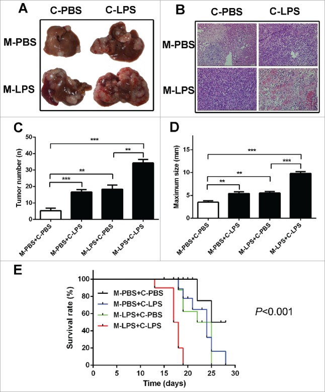
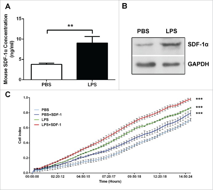
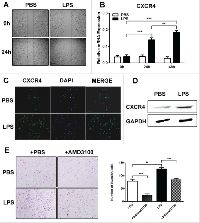
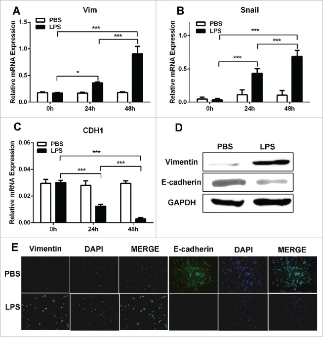
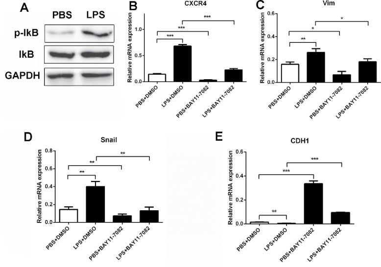
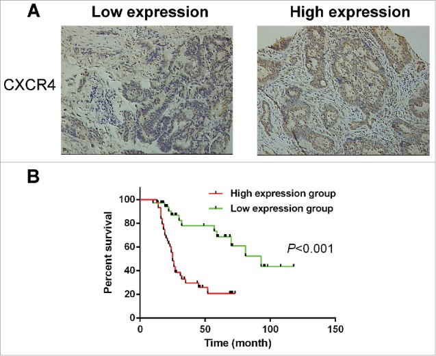
Colorectal cancer (CRC) is the most commonly diagnosed cancer worldwide, and over 50% of patients will develop hepatic metastasis during the course of their disease. CXCR4 and its ligand, stromal cell-derived factor 1α (SDF-1α)/chemokine (C-X-C motif) ligand 12 (CXCL12) have been revealed as regulatory molecules involved in the spreading and progression of a variety of tumors. Here we have shown that lipopolysaccharides (LPS) promoted the migratory capacity of colon cancer cells in vivo and in vitro, which correlated with the activation of SDF-1α/CXCR4 axis and epithelial-mesenchymal transition (EMT) occurrence. Additionally, we found that LPS-induced CXCR4 expression and EMT through NF-κB signaling pathway activation. And inhibition of NF-κB pathway, which recovered the epithelial phenotype and attenuated CXCR4 expression, inhibited cell migratory capacity. Clinically, high levels of CXCR4 always correlated with metastasis and poor prognosis of CRC patients. In conclusion, LPS participate in the whole process of hepatic metastasis of CRC, not only causing liver damage resulting in the production of SDF-1α, but also enhancing the invasive potential of CRC cells by promoting CXCR4 expression and EMT occurrence, which would contribute to the enhancement of cell migration and invasion.

Keywords: NF-κB; SDF-1α/CXCR4 axis; colorectal cancer; epithelial-mesenchymal transition; lipopolysaccharides.


Figures

Figure 1.
Figure 1.
LPS contributed to hepatic metastasis of colon cancer cells. (A) Mouse splenic vein metastasis assay was performed to detect the role of LPS in the hepatic metastasis. (B) H & E staining was performed on serial sections of metastatic tumors and normal liver (× 200). (C) The numbers of nodules were quantified on mice livers (n = 10 per group). Values for individual mice are shown above the bars (**P < 0.01, ***P < 0.001). (D) The maximum size of nodules was quantified on the surface of mice liver. Values for individual mice are shown above the bars (**P < 0.01, ***P < 0.001). (E) The survival time of treated mice were monitored. C-PBS: PBS-challenged C26 cells; C-LPS: LPS-challenged C26 cells; M-PBS: PBS-challenged BALB/c mice; M-LPS: LPS-challenged BALB/c mice.
Figure 2.
Figure 2.
LPS induced SDF-1α expression, which was related to the hepatic metastasis of colon cancer cells. (A) The level of SDF-1α in PBS and LPS-challenged mice was evaluated by ELISA (**P < 0.01). (B) The expression of SDF-1α in mice liver was measured by protein gel blot. (C) Real-time migration assay (xCELLigence System, ACEA Biosciences Inc.) was used to detect the migratory of C26 cells (***P < 0.001).
Figure 3.
Figure 3.
LPS promoted CRC cells migration through upregulating CXCR4 expression in vitro. (A)The wound healing assay was employed to determine the migration of PBS and LPS-disposed C26 cells. Cells were monitored 24 h to evaluate the rate of migration into the scratched area (X40). (B, C and D) RT-PCR, western blot and immunofluorescence were used to detect CXCR4 expression in PBS and LPS-disposed C26 cells, GAPDH was used as an internal reference for RT-PCR and protein gel blot. **P < 0.01, ***P < 0.001. (E) Invasiveness of cells was determined using the Transwell assay (**P < 0.01, ***P < 0.001, X200).
Figure 4.
Figure 4.
EMT contributed to the migratory capacity of LPS-disposed CRC cells. (A, B and C). RT-PCR was used to detect Vimentin, Snail and Ecadherin expression in PBS and LPS-disposed C26 cells, GAPDH was used as an internal reference. *P < 0.05, ***P < 0.001. (D) Western blot was used to detect Vimentin and Ecadherin expression in PBS and LPS-disposed C26 cells, GAPDH was used as an internal reference. (E) Immunofluorescence was used to evaluate the expression of Vimentin and Ecadherin in PBS and LPS-disposed C26 cells.
Figure 5.
Figure 5.
LPS induced CXCR4 expression and EMT through NF-κB pathway activation. (A)Western blot was used to detect the activation of IκB in PBS and LPS-disposed C26 cells, GAPDH was used as an internal reference. (B, C, D and E) CXCR4, Vimentin, Snail and Ecadherin expression were detected by RT-PCR. GAPDH was used as an internal reference. BAY11-7082: IκB inhibitor.
Figure 6.
Figure 6.
High CXCR4 expression correlated with tumor metastasis and poor prognosis. (A) The membranous expression of CXCR4 was seen to be low in 36/80 of CRC tissues (left), and high in 44/80 (right). Magnification: ×200. (B). Overall survival was analyzed in the same cohort of CRC patients and the results showed that CRC patients in the high CXCR4 expression group also have poorer overall survival than those in the low CXCR4 expression group (P < 0.001). CRC: Colorectal cancer.

References

    1. Jemal A, Bray F, Center MM, Ferlay J, Ward E, Forman D. Global cancer statistics. CA Cancer J Clin 2011; 61:69-90; PMID:21296855. - PubMed
    1. Manfredi S, Lepage C, Hatem C, Coatmeur O, Faivre J, Bouvier AM. Epidemiology and management of liver metastases from colorectal cancer. Ann Surg 2006; 244:254-9; PMID:16858188; http://dx.doi.org/ 10.1097/01.sla.0000217629.94941.cf - DOI - PMC - PubMed
    1. Jemal A, Siegel R, Ward E, Hao Y, Xu J, Murray T, Thun MJ. Cancer statistics, 2008. CA Cancer J Clin 2008; 58:71-96; PMID:18287387. - PubMed
    1. Rao R. Endotoxemia and gut barrier dysfunction in alcoholic liver disease. Hepatology 2009; 50:638-44; PMID:19575462; http://dx.doi.org/ 10.1002/hep.23009 - DOI - PMC - PubMed
    1. Rowland IR. The role of the gastrointestinal microbiota in colorectal cancer. Curr Pharm Des 2009; 15:1524-7; PMID:19442169; http://dx.doi.org/ 10.2174/138161209788168191 - DOI - PubMed

Publication types

MeSH terms

LinkOut - more resources