. 2016 Apr;87(4):470-80.
doi: 10.1902/jop.2016.150487. Epub 2016 Jan 8.

Combination of Controlled Release Platelet-Rich Plasma Alginate Beads and Bone Morphogenetic Protein-2 Genetically Modified Mesenchymal Stem Cells for Bone Regeneration

Affiliations

Combination of Controlled Release Platelet-Rich Plasma Alginate Beads and Bone Morphogenetic Protein-2 Genetically Modified Mesenchymal Stem Cells for Bone Regeneration

Gabriela Fernandes et al. J Periodontol. 2016 Apr.

Abstract

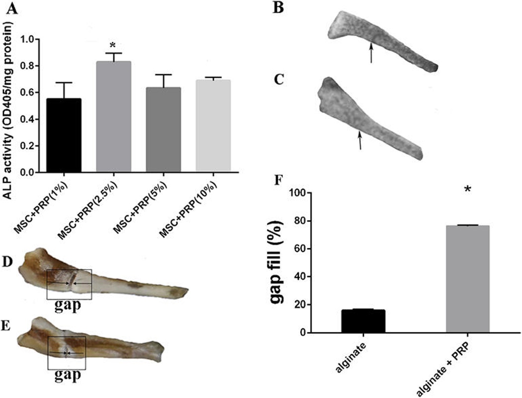
Background: Platelet-rich plasma (PRP) consists of platelet-derived growth factor and transforming growth factor-β that increase proliferation of mesenchymal stem cells (MSCs), whereas bone morphogenetic protein-2 (BMP2) promotes osteogenic differentiation of MSCs. However, the high degradation rate of fibrin leads to the dissociation of cytokines even before the process of bone regeneration begins. To the best of the authors' knowledge, this is the first study to examine the combined effect of sustained release of PRP from alginate beads on BMP2-modified MSC osteogenic differentiation in vitro and sustained release of PRP alone on a fracture defect model ex vivo as well as its effect on calvarial suture closure.

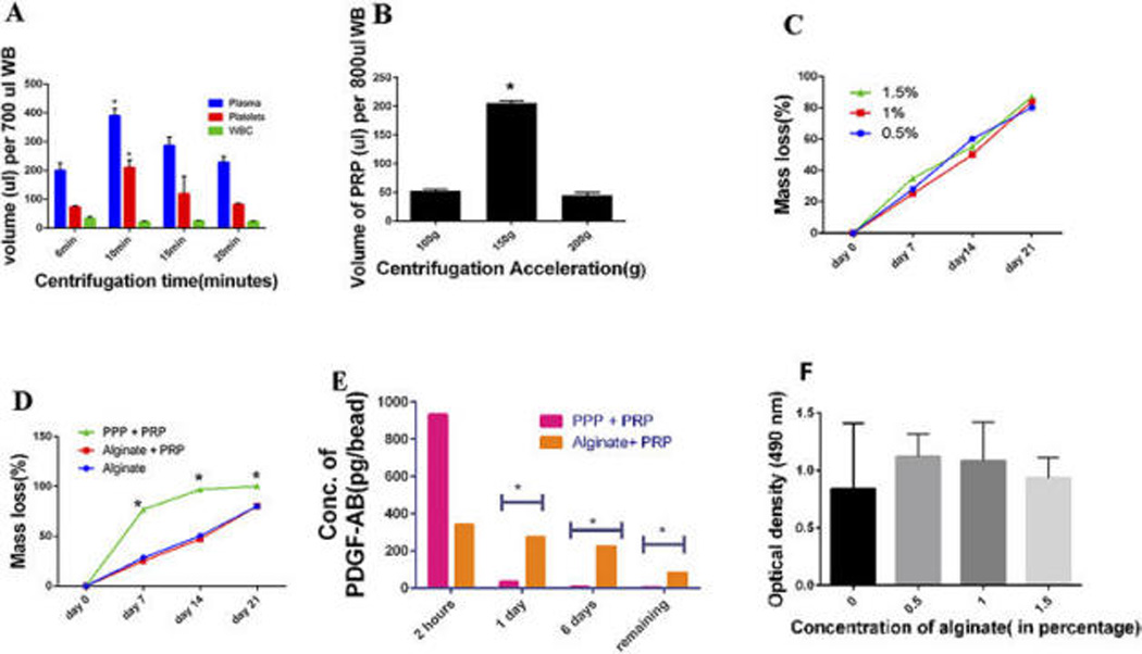
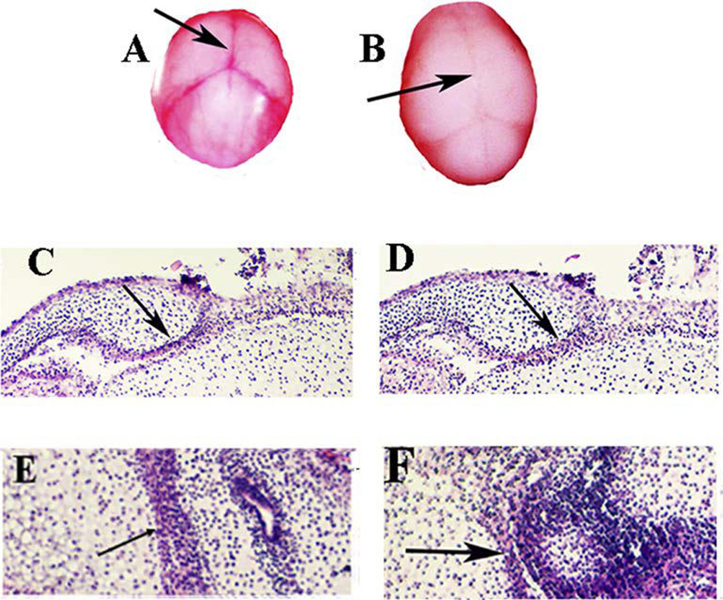
Methods: After optimizing the alginate concentration for microspheres, the combined osteogenic and mineralization effect of PRP and BMP2 on MSCs was studied. Self-setting alginate hydrogel carrying PRP was tested on a femur defect model ex vivo. The effect of PRP at day 15 on the closure of the embryonic mouse calvaria sutures ex vivo was also studied.

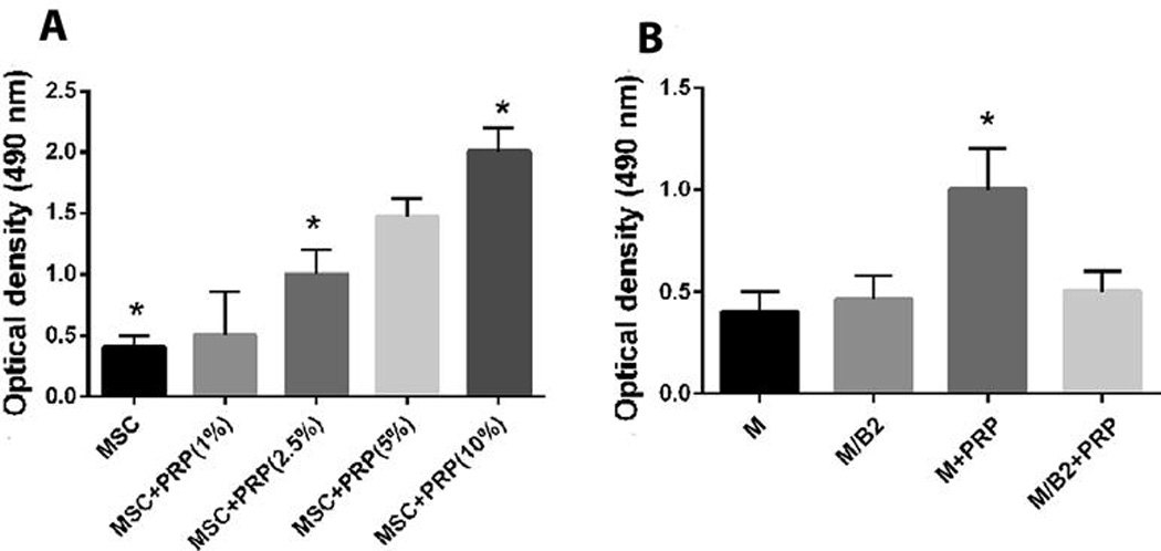
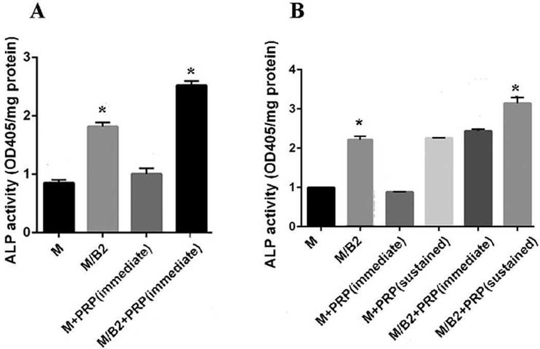
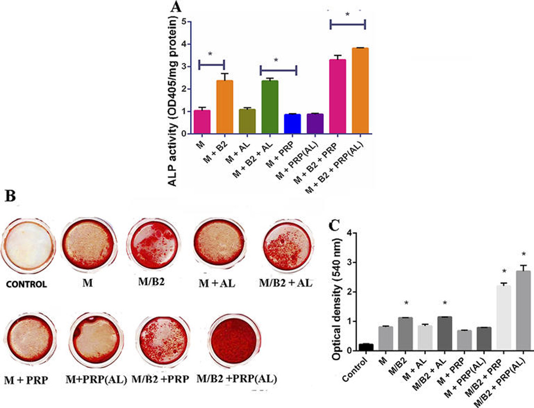
Results: Increase of PRP concentration promoted proliferation of MSCs, and 2.5% to 10% of PRP gradually increased alkaline phosphatase (ALP) activity in the cells in a dose-dependent manner. Sustained release of PRP and BMP2 demonstrated significantly higher ALP and mineralization activity (P <0.05). Radiographs of alginate hydrogel with PRP-treated bone demonstrated nearly complete healing of the fracture, and histologic sections of the embryonic calvaria revealed that PRP leads to suture fusion.

Conclusion: Sustained release of PRP along with BMP2-modified MSCs can significantly promote bone regeneration.

Keywords: Bone regeneration; periodontal regeneration; platelet-rich plasma; stem cells.


Figures

Figure 1
Figure 1
(A) Comparison between the volumes of PPP, PRP, and leucocytes in the upper layer after the first spin step of 150 ×g for 5, 10, 15 and 20 min. Volume of Whole Blood (WB):700 uL. (B) Recovery volume of PRP after the first spin step of WB (800uL) at centrifugal acceleration from 100 to 200 ×g for 10 min (N=3). (C) The formula for mass loss % was calculated as follows: Mass loss % = Original weight – weight after the estimated time period/ Original Weight. The mass loss (%) was assessed for the concentration of 0.5, 1 and 1.5% of alginate. There was no significant difference between the groups (p>0.05). Data represent the mean + SD of N= 3 samples. (D) The degradation rate for the group of alginate microsphere only, Alginate microsphere incorporating PRP and PPP incorporating PRP was assessed at 4 time points. The degradation rate of PPP as compared to Alginate with PRP and alginate alone was significantly higher (p<0.05).There was no difference in the degradation rates of Alginate containing PRP and Alginate alone. Data represent the mean + SD of N= 3 samples. (E) ELISA Assay: The concentration of PDGF was evaluated using an ELISA assay after samples were collected at various time points of 2 hours, 1 day and 6 days and the evaluation of the remaining growth factor where N=3 for the group of Alginate microsphere incorporating PRP and PPP incorporating PRP. PDGF release from the Alginate containing PRP was significantly higher than the PPP + PRP group and demonstrated a consistent release at various time points as compared to the PPP + PRP beads alone. (p<0.05). (F) MTS assay: Optimization of the concentration of the Alginate Microspheres; Effect of the concentration of Alginate Microspheres at 0, 0.5, 1 and 1.5% respectively at a cell seeding density of 4,000 cells/cm2. There was no significant difference in the effect of the various concentrations of alginate on cell viability at 4,000cells/cm2 (p>0.05)
Figure 2
Figure 2
(A) Cell Proliferation activity of the various groups of MSC, MSC + PRP (1 %), MSC + PRP (2.5%), MSC + PRP (5%), MSC + PRP (10%) at a cell seeding density of 1×10⁴ per well in a 96- well plate. Significant difference from the group of MSC vs MSC + PRP (2.5), MSC + PRP(2.5) vs MSC + PRP (10%), MSC vs MSC + PRP(10%) is indicated by * (p <0.01). (B) Cell Proliferation activity of the various groups of MSC, MSC/BMP2, MSC + PRP, MSC/BMP2 + PRP. The cellular proliferation activity of MSC + PRP was significantly higher than the rest of the groups as indicated by * (p<0.01). There is no difference among other three groups.
Figure 3
Figure 3
(A) Groups of MSC, MSC/BMP2, MSC + PRP (immediate) and MSC/BMP2 + PRP (immediate) were evaluated for their ALP Activity on day 2. MSC/BMP2 + PRP (immediate) demonstrated significantly higher ALP activity as compared to the other groups on day 2 (p<0.05). (B) Groups of MSC, MSC/BMP2, MSC + PRP (immediate), MSC+ PRP (sustained), MSC/BMP2 + PRP (immediate) and MSC/BMP2 + PRP (sustained) were evaluated for their ALP Activity on day 7. The group of MSC/BMP2 + PRP (sustained) demonstrated significantly higher ALP activity as compared to other groups* (p<0.05).
Figure 4
Figure 4
ALP Assay and Alizarin red assay: (A) The groups of MSC (M), MSC/BMP2( M/B2), MSC + ALGINATE BEADS(M + AL), MSC/BMP2 + ALGINATE BEADS(M/B2 + AL), MSC + PRP (M + PRP), MSC + PRP(ALGINATE BEADS) (M + PRP(AL)), MSC + BMP2 + PRP (M/B2 + PRP) and MSC/BMP2 + PRP(ALGINATE BEADS) ( M/B2 + PRP(AL)) were assessed for their ALP activity. The cells were induced with OS media for 7 days for ALP assay. Data represent the mean + SD of N= 3 samples. The ALP activity of the group of M/B2 + PRP (AL) was significantly higher than the other groups as marked as * (p<0.05). (B and C) Alizarin red: qualitative and quantitative: Cells were cultured in OS medium with 9 different groups (N=3). Control group: the empty wells were treated with OS medium. The groups of MSC vs MSC/BMP2, MSC vs MSC/BMP2 +ALGINATE, MSC vs MSC/BMP2 + PRP AND MSC vs MSC/BMP2 + PRP(ALGINATE) demonstrated a significant difference (p<0.05) as indicated by * on the graph.
Figure 5
Figure 5
(A) Groups of MSC+PRP (1%), MSC+PRP (2.5%), MSC+ PRP (5%) and MSC + PRP (10%) were assessed for the optimization of PRP concentration. MSC + PRP (2.5%) demonstrated significantly higher ALP activity (N=3) as compared to the other three groups denoted by * (p<0.05). Effect of PRP on a 2 mm mouse femur defect: The gap fill % was calculated using the formula of Gap fill% = Original gap between the bone edges – gap between the bone edges post healing period × 100 Original gap between the bone edges. (B–C) Radiograph depicting complete closure of the defect in the alginate with PRP group (B), whereas the control group still displays the defect (C). (D–E): Images of femur defects with (D) or without PRP treatment (E). (F) Quantitative analysis of gap fill of the defect. 75% of the gap fill of the defect in the group containing alginate gel with PRP and 15% of gap fill was observed in the alginate only group. p<0.05.
Figure 6
Figure 6
Effect of PRP on mouse calvarial suture closure (N=3): (A–B) Calvaria of the mouse were cultured for 5 days for the control group and the group with PRP (150ul) in the OS media (500uL). The group with the PRP shows an approximation of the sutures (Nikon micro VR 85mm) (B), whereas the group without the PRP does not demonstrate any suture closure (A). Histological section (coronal section (C and D) and transverse section (E and F) stained with H & E: The PRP groups showed an approximation of the coronal suture and the sagittal suture (D and F) and the control group failed to do so (C and E).

Similar articles

Cited by

References

    1. Gallini G, Vecchi V, Canzi D. [Bone regeneration and new formation of connective attachment: theory, technic and critical review of the literature] Riv Ital Stomatol. 1984;53(1):5–17. PubMed PMID: 6382563. - PubMed
    1. Mellonig J. Periodontal regeneration with bone grafts. Dent Econ. 1993;83(5):100–101. PubMed PMID: 8243760. - PubMed
    1. Elsalanty ME, Genecov DG. Bone grafts in craniofacial surgery. Craniomaxillofac Trauma Reconstr. 2009;2(3):125–134. PubMed PMID: 22110806; PubMed Central PMCID: PMC3052656. - PMC - PubMed
    1. Zheng L, Wang Q. [The current situation and future of extracellular matrix materials for bone tissue engineering] Sheng Wu Yi Xue Gong Cheng Xue Za Zhi. 2001;18(3):470–474. PubMed PMID: 11605519. - PubMed
    1. Khan Y, Yaszemski MJ, Mikos AG, Laurencin CT. Tissue engineering of bone: material and matrix considerations. J Bone Joint Surg Am. 2008;90(Suppl 1):36–42. PubMed PMID: 18292355. - PubMed

Publication types

Substances