. 2016 Feb;30(2):173-88.
doi: 10.1210/me.2015-1177. Epub 2016 Jan 8.

Structural Analysis on the Pathologic Mutant Glucocorticoid Receptor Ligand-Binding Domains

Affiliations

Structural Analysis on the Pathologic Mutant Glucocorticoid Receptor Ligand-Binding Domains

Darrell E Hurt et al. Mol Endocrinol. 2016 Feb.

Abstract

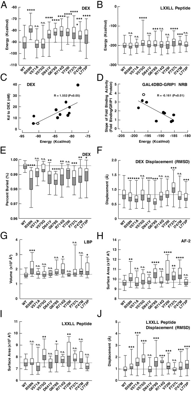
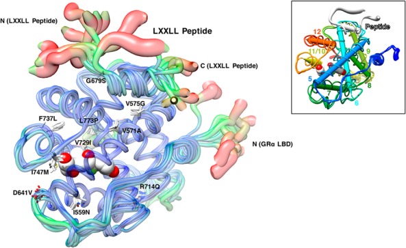
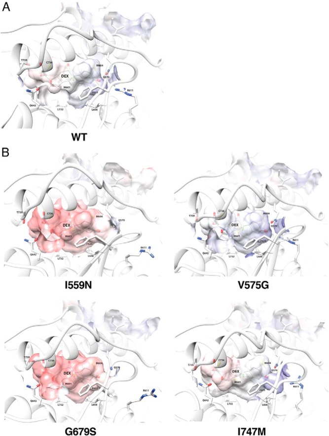
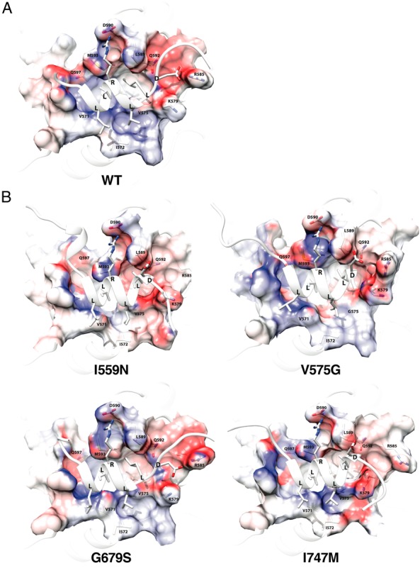
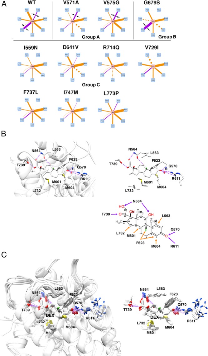
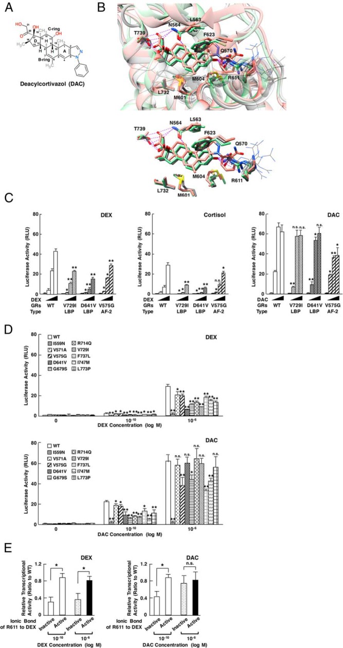
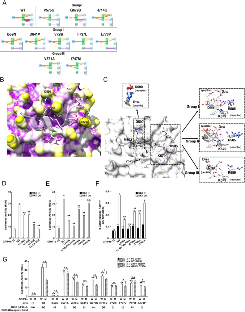
Glucocorticoid receptor (GR) gene mutations may cause familial or sporadic generalized glucocorticoid resistance syndrome. Most of the missense forms distribute in the ligand-binding domain and impair its ligand-binding activity and formation of the activation function (AF)-2 that binds LXXLL motif-containing coactivators. We performed molecular dynamics simulations to ligand-binding domain of pathologic GR mutants to reveal their structural defects. Several calculated parameters including interaction energy for dexamethasone or the LXXLL peptide indicate that destruction of ligand-binding pocket (LBP) is a primary character. Their LBP defects are driven primarily by loss/reduction of the electrostatic interaction formed by R611 and T739 of the receptor to dexamethasone and a subsequent conformational mismatch, which deacylcortivazol resolves with its large phenylpyrazole moiety and efficiently stimulates transcriptional activity of the mutant receptors with LBP defect. Reduced affinity of the LXXLL peptide to AF-2 is caused mainly by disruption of the electrostatic bonds to the noncore leucine residues of this peptide that determine the peptide's specificity to GR, as well as by reduced noncovalent interaction against core leucines and subsequent exposure of the AF-2 surface to solvent. The results reveal molecular defects of pathologic mutant receptors and provide important insights to the actions of wild-type GR.


Figures

Figure 1.
Figure 1.
Pathologic GRα mutants demonstrate changes in the energy required for interacting with dexamethasone (DEX) or the LXXLL peptide, their average displacement over time, the percent buried area (partitioning from the surrounded solvent) of DEX, the volume of LBP, and the surface contacting area of AF-2 and the LXXLL peptide. A, Box plots of the interaction energy for DEX. Most of the mutant receptors increase the energy required for interacting with DEX compared with wild-type (WT) GRα. Mutants V571A and V575G show no statistical difference (n = 200). B, Box plots of the interaction energy for the LXXLL peptide. Mutant receptors V575G, G679S, F737L, and I747M increase the energy required for interacting with the LXXLL peptide (n = 200). C, Correlation between affinity (Kd) of the pathologic GRα mutants to DEX and their calculated energy required for interacting with DEX. Kd values of WT GRα and its pathologic mutants obtained in whole-cell DEX binding assays were compared with their calculated mean energy required for interacting with DEX observed in simulation. Open and closed circles indicate results of the WT GRα and pathologic GRα mutants, respectively. DEX titration curves for bound radioactive DEX and subsequent Scatchard plots for WT GRα and its mutant receptors are shown in Supplemental Figure 2. D, Correlation between slopes of the fold binding activity of WT GRα and its mutant receptors to GRIP1 NRB obtained in the mammalian 2-hybrid assay and their calculated energy required for interacting with the LXXLL peptide. Mammalian 2-hybrid assays employing WT GRα or mutant receptor LBD fused with GAL4 DBD and grading amounts of the GRIP1 NRB fused with VP16 AD in the presence or absence of 10−5M DEX were performed, and slopes of their fold binding activity obtained with linear regression analysis (Supplemental Figure 3A) were compared with their calculated mean energy required for interacting with the LXXLL peptide observed in simulation. Open and closed circles indicate results of the WT GRα and pathologic GRα mutants, respectively. Note that the levels of expressed VP16 AD-fused GRIP1 NRB are almost linear in the range of its transfected plasmid amounts employed in this assay (Supplemental Figure 3B). Similar expression of GAL4DBD-GRα LBDs is shown in Supplemental Figure 1B. Representative results of the mammalian 2-hybrid assay using GAL4DBD-GRα LBD (WT) are shown in Supplemental Figure 3C. E, Box plots of the percent buried area of DEX. All pathologic GRα mutants except V571V, V575G, and V729I have significantly less buried area of DEX than WT GRα (n = 20). F, Box plots of the DEX displacement shown with RMSD. Mutant receptors I559N, V571A, D641V, G679S, and V773P have significantly less distance to DEX than WT GRα, whereas F737L has increased distance (n = 20). G, Box plots of the LBP volume. Mutant receptors I559N, R714Q, and L773P have significantly larger LBP volume than WT GRα, suggesting a less snug fit to DEX (n = 20). H and I, Box plots of the surface contacting area of AF-2 and the LXXLL peptide. Mutant receptors I559N, G679S, V729I, F737L, and I747 have significantly more surface contacting area of their AF-2 to the LXXLL peptide than WT GRα (H), whereas the mutant receptors, V575G, G689S, and V729I, have more contacting surface area of the LXXLL peptide to their AF-2 (I) (n = 20). J, Box plots of the LXXLL peptide displacement shown with RMSD. Mutant receptors I559N, V571A, V575G, G679S, R714Q, V729I, I747M, and L773P have significantly more distance to the LXXLL peptide than wild-type GRα (n = 20). Box plots of each panel illustrate the medians, and upper and lower quartiles, whereas the whiskers indicate range of distribution. The median value of WT GRα in each analysis is indicated with a dotted line. *, P < .05; **, P < .01; ***, P < .001; ****, P < .001; n.s., not significant, compared with WT GRα. WT, wild-type.
Figure 2.
Figure 2.
Averaged trajectories reveal slight changes in overall conformation of pathologic GRα mutant LBDs and their interaction with the LXXLL peptide. Thickness and color of the overlaid Cα-traces of mutant receptor LBDs indicate the areas of least (thin and blue) to most (thick and red) motion over the course of simulation. Locations and side chains of the mutated amino acids are indicated, whereas dexamethasone (shown with the white and red spheres of space-filling model) is located inside LBP. In the right upper inset, helix-5 to helix-12 of the wild-type GRα LBD are shown with colored dipoles. The movie rotating this image is available as Supplemental Movie 1.
Figure 3.
Figure 3.
Four representative pathologic GRα mutants change conformational and chemical properties of their LBP. LBP of the wild-type GRα (A) and 4 representative pathologic GRα mutants (I559N, ligand-binding defective; V575G, AF-2 defective; G679S and I747M, both defective) (B) are shown. Positive and negative electrostatic potential are indicated with blue and red, respectively. The key residues of the receptors making important molecular interactions to dexamethasone are incorporated. The structures shown and their calculated biochemical properties are those of the averaged trajectories. DEX, dexamethasone; WT, wild type.
Figure 4.
Figure 4.
Four representative pathologic GRα mutants change conformational and chemical properties of their AF-2 surface. The AF-2 surface of wild-type GRα (A) and 4 representative pathologic GRα mutants (I559N, ligand-binding defective; V575G, AF-2 defective; G679S and I747M, both defective) (B) are shown. Positive and negative electrostatic potential are indicated with blue and red, respectively. Key residues of the receptors and the LXXLL peptide that make important molecular interactions are incorporated. The structures shown and their calculated biochemical properties are those of the averaged trajectories. WT, wild type.
Figure 5.
Figure 5.
Alteration of the electrostatic bond formed by R611 and T739 of pathologic GRα mutants may largely explain the reduced affinity of many pathologic GRα mutants to this steroid. A, Diagram of individual molecular interactions between dexamethasone and wild-type (WT) GRα or pathologic GRα mutants. Purple and orange lines indicate electrostatic and noncovalent bonds, respectively. Line thickness indicates frequency of the observed interaction (thus strength of interaction), whereas dotted lines indicate no statistical change in the frequency of interaction compared with WT GRα. Only the interactions dramatically altered in the mutant receptors are shown. Arrows are for attracting readers' attention. Based on the strength of interaction created by R611 and T739 of receptors, pathologic GRα mutants can be categorized into 3 groups (groups A, B, and C). Full interaction diagrams are shown in Supplemental Figure 4. B, 3D or schematic models (residues only) of the molecular interaction between WT GRα LBP and dexamethasone. Top left and top right panels demonstrate 3D interaction images of dexamethasone and the key residues of WT GRα that create important contacts to this steroid. Bottom right panel shows schematic molecular interaction between WT GRα and dexamethasone. Purple and orange arrows indicate electrostatic and noncovalent bonds, respectively. C, Superimposed 3D images of dexamethasone and the key residues of all pathologic GRα mutants. Panels demonstrate superimposed 3D interaction images of dexamethasone and the key residues of all pathologic GRα mutants. Among the key amino acids of pathologic mutants participating in interaction with dexamethasone, R611 is largely deviated in these mutant receptors, which underlies reduced/disappeared electrostatic interaction between this residue and carbonyl oxygen at carbon-3 of dexamethasone. Q570 and N564 are omitted from these panels. DEX, dexamethasone; WT, wild type.
Figure 6.
Figure 6.
Deacylcortivazol (DAC) is a more potent agonist for the pathologic GRα mutants with defective LBP than dexamethasone (DEX) by filling the space created by deviated R611 with its bulky phenylpyrazole structure. A, Molecular structure model of DAC. DAC has a large phenylpyrazole group at its steroid A-ring. B, Superimposed 3D LBP models of wild-type (WT) GRα bound with DEX or DAC. 3D image of WT GRα LBP bound with DEX or DAC are shown (white, the reported crystallographic image with DEX [PDB ID, 1M2Z]; green, our simulation image associated with DEX; and orange, the reported crystallographic image with DAC [PDB ID, 3BQD]). Note that the former 2 images highly overlap with each other. Cα anchor position of R611 does not change between the green and the orange image, while its side chain significantly shifts to the similar direction as those observed in pathologic GRα mutants (shown with thin gray lines, obtained with the simulation for the mutant LBDs associated with DEX), forming a space filled with a large phenylpyrazole group of this compound. Electrostatic interactions of DEX or DAC to GRα residues are shown with purple broken lines. C, DEX, cortisol, and DAC differentially stimulate the transcriptional activity of GRα V729I, D641V, and V575G. HCT116 cells were transfected with WT or indicated mutant GRα-expressing plasmid together with pOLDO-MMTV-Luc and pGL4.73[hRluc/SV40] in the presence of 0M, 10−10M, 10−8M, or 10−6M DEX (left panel), cortisol (middle panel), or DAC (right panel). Bars represent mean ± SE values of firefly luciferase activity normalized for Renilla luciferase activity. WT, wild type. *, P < .05; **, P < .01; n.s., not significant, compared with the values of WT GRα treated with the same concentrations of compound (n = 3). D, DEX and DAC differentially stimulates the transcriptional activity of pathologic GRα mutants at its 2 concentrations. HCT116 cells were transfected with WT or indicated mutant GRα-expressing plasmid together with pOLDO-MMTV-Luc and pGL4.73[hRluc/SV40] in the presence of 0M, 10−10M, or 10−6M DEX (top panel) or DAC (bottom panel). Bars represent mean ± SE values of firefly luciferase activity normalized for Renilla luciferase activity. WT, wild type. *, P < .05; **, P < .01; n.s., not significant, compared with the values of WT GRα treated with the same concentrations of compound (n = 3). E, DAC improves the transcriptional activity of pathologic GRα mutants in contrast to DEX. Pathologic GRα mutants and WT GRα are categorized into 2 groups, based on their property of the electrostatic bond at R611: no or weak electrostatic bond (inactive) (<30% of the WT GRα): I559N, D641V, G679S, V729I, F737L, and I747M; and active bond (active) (>90% of the WT GRα): V571A, V575G, and WT GRα. Their relative transcriptional activity against WT GRα at 10−10M or 10−6M DEX (left panel) or DAC (right panel) was then compared. Bars represent mean ± SE values of fold transcriptional activity of GRαs (firefly luciferase activity normalized for Renilla luciferase activity) against that of the WT GRα. WT, wild type. *, P < .05; n.s., not significant, compared with the conditions indicated (inactive group, n = 6 and active group, n = 3).
Figure 7.
Figure 7.
Loss/reduction of the electrostatic bond between R585 of pathologic GRα mutants and noncore LXXLL residues (especially D750) of the LXXLL peptide contributes to the reduced AF-2 activity of these mutant receptors. A, Diagram of individual molecular interactions between the LXXLL peptide and wild-type (WT) GRα or pathologic GRα mutants. Panels are created by following the same rules as that employed in Figure 5A. For model demonstration, tyrosine (Y) located 1 amino acid N-terminally to the third core leucine is omitted, as its side chain protrudes into free space opposite to the AF-2 surface and it does not make any interactions with AF-2. All mutant receptors can be categorized into 3 groups based on the 2 interactions indicated with brackets (groups I, II, and III). Full interaction diagrams are shown in Supplemental Figure 5. B, 3D interaction images of WT GRα AF-2 and the LXXLL peptide. The AF-2 surface of WT GRα has 3 large pockets into which core leucines (L745, L748, and L749) of the LXXLL peptide deeply bury themselves. There are additional intermolecular contacts that are important for peptide binding, including the electrostatic bonds created between 1) R746 (LXXLL peptide) and D590 (receptor), 2) D750 (LXXLL peptide) and R585 (receptor), and 3) D752 (LXXLL peptide) and K579 (receptor). C, Deviation of the receptor residues forming electrostatic bonds against noncore LXXLL residues. 3D image indicates molecular interaction between the LXXLL peptide (peptide) and key residues of WT GRα. The LXXLL peptide forms important electrostatic bonds with its noncore leucine residues (R746, D750, and D752) against the receptor residues (D590, R585, and K579, respectively). Pathologic GRα mutants demonstrated significant shift of the side chains of some of these receptor residues, among which the side chain of R585 demonstrated the most significant deviation. Molecular interaction and side chain shift of groups I, II, and III receptors are shown in right insets. Top left inset contains the minimally deviated side chains of D590 (receptor) and their interacting partner R746 (LXXLL peptide). D, The third (C-terminal) LXXLL motif of GRIP1 NRB contributes most significantly to the enhancement of GRα transcriptional activity by this coactivator. HCT116 cells were transfected with indicated GRIP1-expressing plasmids together with WT GRα-expressing plasmid, pMMTV-Luc and pGL4.73[hRluc/SV40], and were incubated in the presence or absence of 10−6M dexamethasone (DEX). Bars represent mean ± SE values of firefly luciferase activity normalized for Renilla luciferase activity. L Mut, GRIPI mutant defective in indicated LXXLL motif(s). **, P < .01, compared with the condition with WT GRIP1 and DEX (n = 3). E, R746 and D750, but not D752, of GRIP1 contributes to the enhancement of GRα transcriptional activity by this coactivator. HCT116 cells were transfected with indicated GRIP1-expressing plasmids together with WT GRα-expressing plasmid, pMMTV-Luc and pGL4.73[hRluc/SV40], and were incubated in the presence or absence of 10−6M DEX. Bars represent mean ± SE values of firefly luciferase activity normalized for Renilla luciferase activity. **, P < .01; n.s., not significant, compared with the condition with WT GRIP1 and DEX (n = 3). F, R746, D750, and D752 of GRIP1 contribute to the binding of GRIP1 NRB to WT GRα LBD in a yeast 2-hybrid assay. EGY48 yeast cells were transformed with p8Op-LacZ, pLexA-GRα (480–777) and indicated GRIP1 NRB-expressing pB42AD-derived plasmids. Bars represent mean ± SE values of β-galactosidase activity corrected for cell density. *, P < .05; **, P < .01, compared with the condition with WT GRIP1 NRB and DEX (n = 3). G, Damaged electrostatic bond of some pathologic GRα mutants to GRIP1 D750 underlies the defective enhancement of their transcriptional activity by GRIP1. HCT116 cells were transfected with WT GRIP1- or GRIP1 D750A-expressing plasmids together with WT GRα- or its mutant receptor-expressing plasmid, pMMTV-Luc and pGL4.73[hRluc/SV40], and were incubated in the presence or absence of 10−6M DEX. Bars represent mean ± SE values of firefly luciferase activity normalized for Renilla luciferase activity. Presence or absence of the electrostatic bond between D750 (LXXLL) and R585 (receptor) are demonstrated under x-axis. *, P < .05; **, P < .01; n.s., not significant, compared with the condition in the presence of WT GRIP1 and DEX, or the 2 conditions indicated (n = 3).

References

    1. Chrousos GP. Stress and disorders of the stress system. Nat Rev Endocrinol. 2009;5:374–381. - PubMed
    1. Kino T, Chrousos GP. Glucocorticoid effect on gene expression. In: Steckler T, Kalin NH, Reul JMHM, eds. Handbook on Stress and the Brain. Amsterdam, The Netherlands: Elsevier BV; 2005:295–312.
    1. Chrousos GP, Kino T. Intracellular glucocorticoid signaling: a formerly simple system turns stochastic. Sci STKE. 2005;2005:pe48. - PubMed
    1. Kino T. Glucocorticoid receptor. In: Chrousos GP, ed. Adrenal Disease and Function. South Dartmouth, MA: Endotext; 2000.
    1. Kino T, Su YA, Chrousos GP. Human glucocorticoid receptor isoform β: recent understanding of its potential implications in physiology and pathophysiology. Cell Mol Life Sci. 2009;66:3435–3448. - PMC - PubMed

Publication types

MeSH terms