. 2016 Jan 8;11(1):e0146460.
doi: 10.1371/journal.pone.0146460. eCollection 2016.

Mutations of Arabidopsis TBL32 and TBL33 Affect Xylan Acetylation and Secondary Wall Deposition

Affiliations

Mutations of Arabidopsis TBL32 and TBL33 Affect Xylan Acetylation and Secondary Wall Deposition

Youxi Yuan et al. PLoS One. .

Abstract

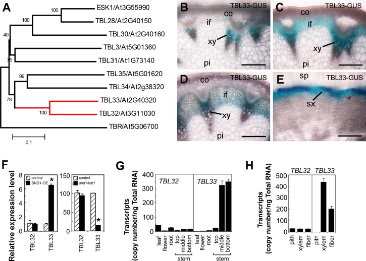
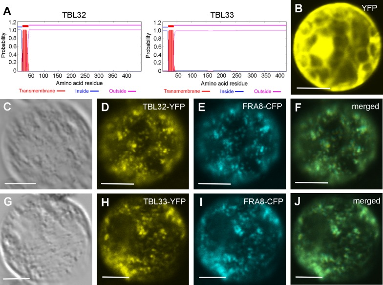
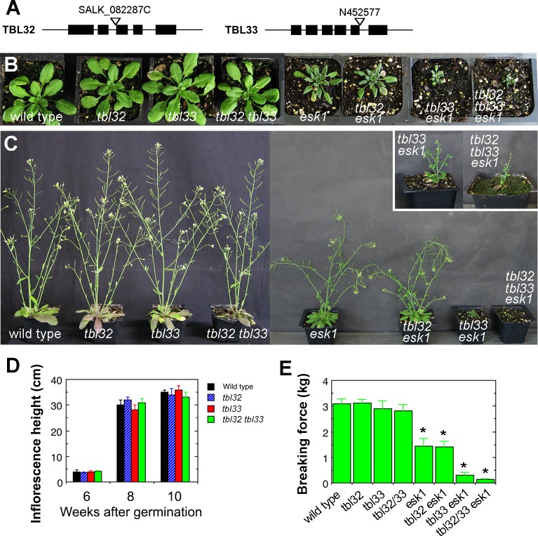
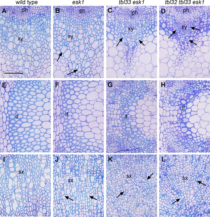
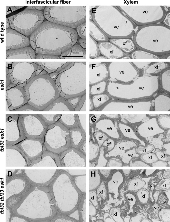
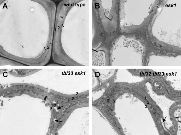
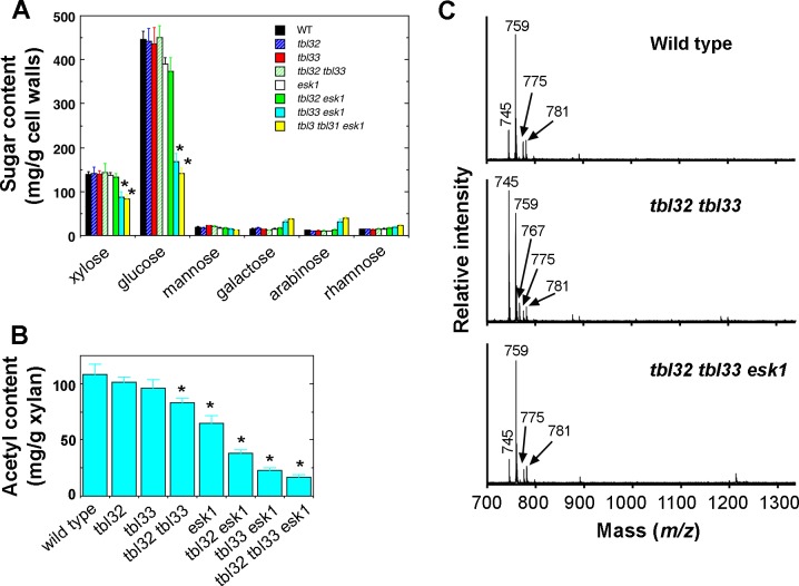
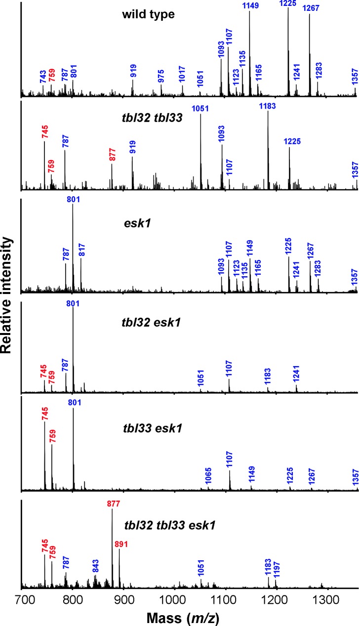
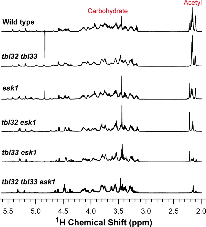
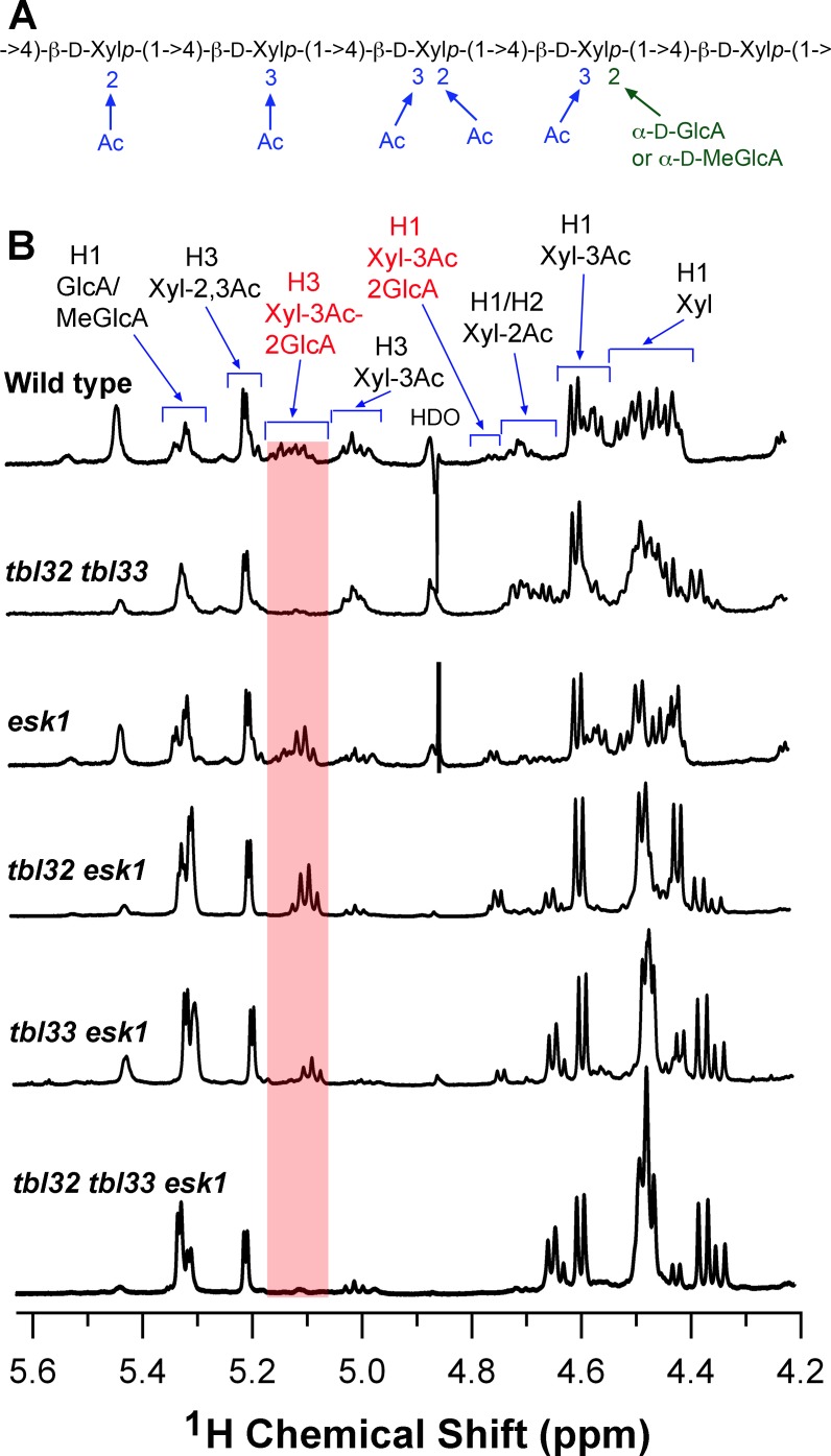
Xylan is a major acetylated polymer in plant lignocellulosic biomass and it can be mono- and di-acetylated at O-2 and O-3 as well as mono-acetylated at O-3 of xylosyl residues that is substituted with glucuronic acid (GlcA) at O-2. Based on the finding that ESK1, an Arabidopsis thaliana DUF231 protein, specifically mediates xylan 2-O- and 3-O-monoacetylation, we previously proposed that different acetyltransferase activities are required for regiospecific acetyl substitutions of xylan. Here, we demonstrate the functional roles of TBL32 and TBL33, two ESK1 close homologs, in acetyl substitutions of xylan. Simultaneous mutations of TBL32 and TBL33 resulted in a significant reduction in xylan acetyl content and endoxylanase digestion of the mutant xylan released GlcA-substituted xylooligomers without acetyl groups. Structural analysis of xylan revealed that the tbl32 tbl33 mutant had a nearly complete loss of 3-O-acetylated, 2-O-GlcA-substituted xylosyl residues. A reduction in 3-O-monoacetylated and 2,3-di-O-acetylated xylosyl residues was also observed. Simultaneous mutations of TBL32, TBL33 and ESK1 resulted in a severe reduction in xylan acetyl level down to 15% of that of the wild type, and concomitantly, severely collapsed vessels and stunted plant growth. In particular, the S2 layer of secondary walls in xylem vessels of tbl33 esk1 and tbl32 tbl33 esk1 exhibited an altered structure, indicating abnormal assembly of secondary wall polymers. These results demonstrate that TBL32 and TBL33 play an important role in xylan acetylation and normal deposition of secondary walls.


Conflict of interest statement

Competing Interests: The authors have declared that no competing interests exist.

Figures

Fig 1
Fig 1. Expression analysis of the Arabidopsis TBL32 and TBL33 genes.
(A) Phylogenetic relationship of TBL32, TBL33, and other ESK1 homologs. TBR, another member of the Arabidopsis DUF231 family, is also included in the phylogenetic analysis. The phylogenetic tree was constructed with the neighbor-joining algorithm. The 0.1 scale denotes 10% change, and the bootstrap values resulted from 1,000 replicates are presented in percentages at the nodes. (B) to (E) Expression patterns of TBL33 in Arabidopsis inflorescence stems and root-hypocotyls. Cross sections of the top stem (C), middle stem (D), bottom stem (E), and root-hypocotyl (F) of TBL33::TBL33-GUS plants were stained for GUS activity (blue). co, cortex; if, interfascicular fiber; pi, pith; sp, secondary phloem; sx, secondary xylem; xy, xylem. Bars = 165 μm. (F) Quantitative PCR analysis of the expression of TBL32 and TBL33 in the SND1 overexpressors (SND1-OE) (left panel) and the snd1 nst1 double mutant (right panel). The expression of each gene in the wild type was used as a control by setting to 1 for SND1 overexpressors and by setting to 100 for the snd1 nst1 double mutant. Asterisks indicate statistically significant differences compared with the control (p < 0.001). (G) Quantitative PCR analysis of the expression of TBL32 and TBL33 in different Arabidopsis organs. (H) Quantitative PCR analysis of the expression of TBL32 and TBL33 in pith, xylem and interfascicular fibers in wild-type stems. The expression level in (G) and (H) was presented as transcript copy numbers per ng total RNA. Error bars denote SD of three biological replicates for each sample.
Fig 2
Fig 2. TBL32 and TBL33 are co-localized with FRA8.
Arabidopsis leaf protoplasts expressing fluorescent protein-tagged fusion proteins were visualized for fluorescent signals with a laser confocal microscope. (A) TBL32 and TBL33 are type II membrane proteins based on the TMHMM2.0 program. Outside, the noncytoplasmic side of the membrane (Golgi); inside, the cytoplasmic side of the membrane. (B) An Arabidopsis protoplast expressing YFP alone showed the distribution of the signals throughout the cytoplasm. (C) to (F) An Arabidopsis protoplast (C) co-expressing TBL32-YFP (D) and the Golgi-localized FRA8-CFP (E). Note the overlap of the signals of TBL32-YFP and FRA8-CFP (F). (G) to (J) An Arabidopsis protoplast (G) co-expressing TBL33-YFP (H) and FRA8-CFP (I). Note the overlap of the signals of TBL33-YFP and FRA8-CFP (J). Bars in (B) to (J) = 12 μm.
Fig 3
Fig 3. Effects of mutations of TBL32, TBL33, and ESK1 on stem mechanical strength and plant growth.
(A) The sites of T-DNA insertions in the TBL32 and TBL33 genes. Filled boxes represent exons. (B) Morphology of 4-week-old seedlings of the wild type and various mutants. (C) Morphology of 8-week-old wild-type and mutant plants. Inset shows the images of a 12-week-old plant of tbl33 esk1 and a 16-week-old plant of tbl32 tbl33 esk1. Note the extremely dwarfed plants of tbl33 esk1 and tbl32 tbl33 esk1. (D) Measurement of inflorescence stem heights during different developmental stages in the wild type, tbl32, tbl33, and tbl32 tbl33 mutants. (E) Stem strength measurement in the wild type and various mutants. Basal parts of mature stems were measured for their breaking force. Error bars in (D) and (E) are SD of measurements from 20 independent plants. Asterisks in (E) indicate statistically significant differences compared with the wild type (p < 0.001).
Fig 4
Fig 4. Effects of mutations of TBL32, TBL33, and ESK1 on vessel morphology and secondary wall thickening.
The bottom internodes of stems and the root-hypocotyls of 8-week-old wild-type and esk1 plants, 12-week-old tbl33 esk1 plants, and 16-week-old tbl32 tbl33 esk1 plants were sectioned for visualization of anatomical structures. (A) to (D) Cross sections of stem xylem bundles showing vessels (arrows) with a mild deformation in esk1 (B) and a severe deformation in tbl33 esk1 (C) and tbl32 tbl33 esk1 (D) compared with the wild type (A). (E) to (H) Cross sections of stem interfascicular regions showing defective secondary wall thickening in esk1 (F), tbl33 esk1 (G), and tbl32 tbl33 esk1 (H) compared with the wild type (E). (I) to (L) Cross sections of root-hypocotyls showing vessels (arrows) with a mild deformation in esk1 (J) and a severe deformation in tbl33 esk1 (K) and tbl32 tbl33 esk1 (L) compared with the wild type (I). if, interfascicular fiber; ph, phloem; sx, secondary xylem; xy, xylem. Bar in (A) = 68 μm for (A) to (L).
Fig 5
Fig 5. Transmission electron micrographs of interfascicular fibers and xylem in the wild type and various mutants.
The bottom internodes of stems of 8-week-old wild-type and esk1 plants, 12-week-old tbl33 esk1 plants, and 16-week-old tbl32 tbl33 esk1 plants were sectioned for visualization of walls of interfascicular fibers and xylem vessels. (A) to (D) Cross sections of interfascicular fibers showing defective secondary wall thickening in esk1 (B), tbl33 esk1 (C), and tbl32 tbl33 esk1 (D) compared with the wild type (A). (E) to (H) Cross sections of xylem cells showing various degrees of deformation in vessels in esk1 (F), tbl33 esk1 (G), and tbl32 tbl33 esk1 (H) compared with the wild type (E). ve, vessel; xf, xylary fiber. Bar in (A) = 10.5 μm for (A) to (H).
Fig 6
Fig 6. Effects of reduced acetylation of xylan on secondary wall structure in xylem vessels of esk1 (B), tbl33 esk1 (C) and tbl32 tbl33 esk1 (D) compared with the wild type (A).
Ultrathin stem sections were stained with lead citrate and uranyl acetate and visualized for vessel secondary wall structure under a transmission electron microscope. The numbers 1, 2 and 3 marked on vessel secondary walls denote the S1, S2 and S3 layers, respectively. Note the drastic alteration in the staining pattern of the S2 layer as well as disintegrated walls in the S2 layer (arrows) in tbl33 esk1 (C) and tbl32 tbl33 esk1 (D). Bar in (A) = 3 μm for (A) to (D).
Fig 7
Fig 7. Measurement of cell wall sugar composition and acetyl contents in the wild type and various mutants.
The inflorescence stems of 8-week-old wild type and esk1 plants, 12-week-old tbl33 esk1 plants, and 16-week-old tbl32 tbl33 esk1 plants were used for extraction of cell wall residues and xylan. (A) Cell wall composition analysis revealed a reduction in the amounts of xylose and glucose in tbl33 esk1 and tbl32 tbl33 esk1 compared with the wild type and esk1. (B) Acetyl contents in DMSO-extracted xylans of the wild type and various mutants. Note the drastic reduction in the acetyl contents in tbl32 esk1, tbl33 esk1, and tbl32 tbl33 esk1. Error bars denote SD of the data from three separate pools of samples. Asterisks in (A) and (B) indicate statistically significant differences compared with the wild type (p < 0.001). (C) MALDI-TOF-MS analysis of xylooligomers generated by endoxylanase digestion of KOH-extracted xylan from the wild type (top panel), tbl32 tbl33 (middle panel) and tbl32 tbl33 esk1 (bottom panel). The ion peaks at m/z 745 and 759 are attributed to (GlcA)Xyl4 and (MeGlcA)Xyl4, respectively. Those at m/z 767 and 781 correspond to the disodiated species of (GlcA)Xyl4 and (MeGlcA)Xyl4, respectively. The ion at m/z 775 corresponds to (Gal-GlcA)Xyl3 [38].
Fig 8
Fig 8. MALDI-TOF MS analysis of acetylated xylans of the wild type and various mutants.
DMSO-extracted xylans were digested with endoxylanase and subject to MALDI-TOF MS. The major ion peaks of masses are indicated and their xylooligomer structures are listed below. Xyln(GlcA)n(Ac)n denotes a xylooligomer (with n number of xylosyl residues) substituted with n number of GlcA and n number of acetyl groups. m/z 743, Xyl5(Ac); m/z 745, Xyl4(GlcA); m/z 759, Xyl4(MeGlcA); m/z 787, Xyl4(GlcA)(Ac); m/z 801, Xyl4(MeGlcA)(Ac); m/z 817, Xyl3(MeGlcA)2; m/z 843, Xyl4(MeGlcA)(Ac)2; m/z 877, Xyl5(GlcA); m/z 891, Xyl5(MeGlcA); m/z 919, Xyl5(GlcA)(Ac); m/z 975, Xyl5(MeGlcA)(Ac)2; m/z 1017, Xyl5(MeGlcA)(Ac)3; m/z 1051, Xyl6(GlcA)(Ac); m/z 1065, Xyl6(MeGlcA)(Ac); m/z 1093; Xyl6(GlcA)(Ac)2; m/z 1107; Xyl6(MeGlcA)(Ac)2; m/z 1123, Xyl5(MeGlcA)2(Ac); m/z 1135, Xyl6(GlcA)(Ac)3; m/z 1149, Xyl6(MeGlcA)(Ac)3; m/z 1165, Xyl5(MeGlcA)2(Ac)2; m/z 1183, Xyl7(GlcA)(Ac); m/z 1197, Xyl7(MeGlcA)(Ac); m/z 1225, Xyl7(GlcA)(Ac)2; m/z 1241, Xyl6(GlcA)(MeGlcA)(Ac); m/z 1267, Xyl7(GlcA)(Ac)3; m/z 1283, Xyl6(GlcA)(MeGlcA)(Ac)2; m/z 1357, Xyl8(GlcA)(Ac)2. Note the increase in the abundance of ions at m/z 745, 759, 877, and 891 (marked in red) corresponding to non-acetylated xylooligomers in tbl32 tbl33, tbl32 esk1, tbl33 esk1, and tbl32 tbl33 esk1.
Fig 9
Fig 9. 1H NMR spectra of acetylated xylans from the wild type, tbl32 tbl33, esk1, tbl32 esk1, tbl33 esk1, and tbl32 tbl33 esk1.
The resonance regions corresponding to acetyl groups and carbohydrate are indicated.
Fig 10
Fig 10. Distribution patterns of xylan acetyl substitutions in the wild type and various mutants.
(A) Diagram of an acetylated xylooligomer from wild-type Arabidopsis xylan. (B) The fingerprint regions of the 1H NMR spectra of acetylated xylans from the wild type, tbl32 tbl33, esk1, tbl32 esk1, tbl33 esk1, and tbl32 tbl33 esk1. The resonances for non-acetylated (Xyl), 2-O-acetylated (Xyl-2Ac), 3-O-acetylated (Xyl-3Ac), 2,3-di-O-acetylated (Xyl-2,3Ac), 3-O-acetylated 2-O-GlcA-substituted xylosyl residues (Xyl-3Ac-2GlcA) and GlcA/MeGlcA are labeled. Note the loss of the resonances of Xyl-3Ac-2GlcA in tbl32 tbl33 and tbl32 tbl33 esk1.

References

    1. Gille S, Pauly M (2012) O-acetylation of plant cell wall polysaccharides. Front Plant Sci 3: 12 10.3389/fpls.2012.00012 - DOI - PMC - PubMed
    1. Pawar PM-A, Koutaniemi S, Tenkanen M, Mellerowicz EJ (2013) Acetylation of woody lignocellulose: significance and regulation. Front Plant Sci 4: 118 10.3389/fpls.2013.00118 - DOI - PMC - PubMed
    1. Yuan Y, Teng Q, Zhong R, Ye Z-H (2013) The Arabidopsis DUF231 domain-containing protein ESK1 mediates 2-O- and 3-O-acetylation of xylosyl residues in xylan. Plant Cell Physiol 54: 1186–1199. 10.1093/pcp/pct070 - DOI - PubMed
    1. Xiong G, Cheng K, Pauly M (2013) Xylan O-acetylation impacts xylem development and enzymatic recalcitrance as indicated by the Arabidopsis mutant tbl29. Mol Plant 6: 1373–1375. 10.1093/mp/sst014 - DOI - PubMed
    1. Gou JY, Miller LM, Hou G, Yu XH, Chen XY, Liu CJ (2012) Acetylesterase-mediated deacetylation of pectin impairs cell elongation, pollen germination, and plant reproduction. Plant Cell 24: 50–65. 10.1105/tpc.111.092411 - DOI - PMC - PubMed

Publication types