

A Shh-Foxf-Fgf18-Shh Molecular Circuit Regulating Palate Development
- PMID: 26745863
- PMCID: PMC4712829
- DOI: 10.1371/journal.pgen.1005769
A Shh-Foxf-Fgf18-Shh Molecular Circuit Regulating Palate Development
Abstract
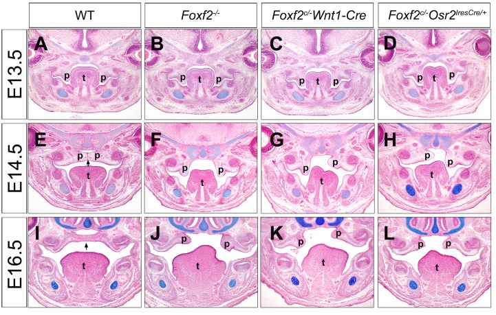
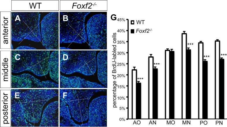
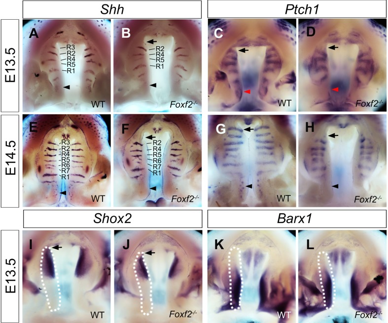
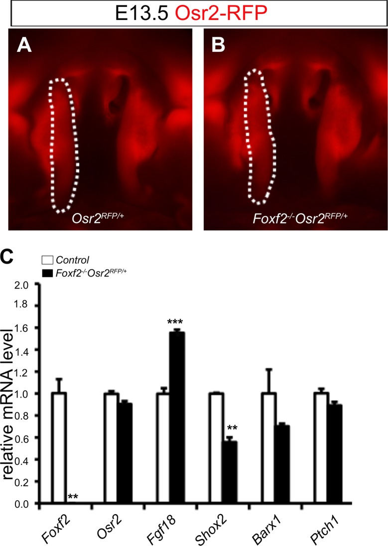
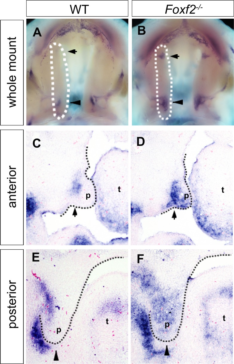
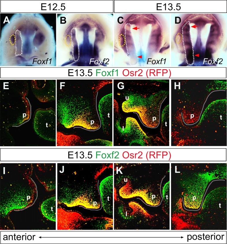
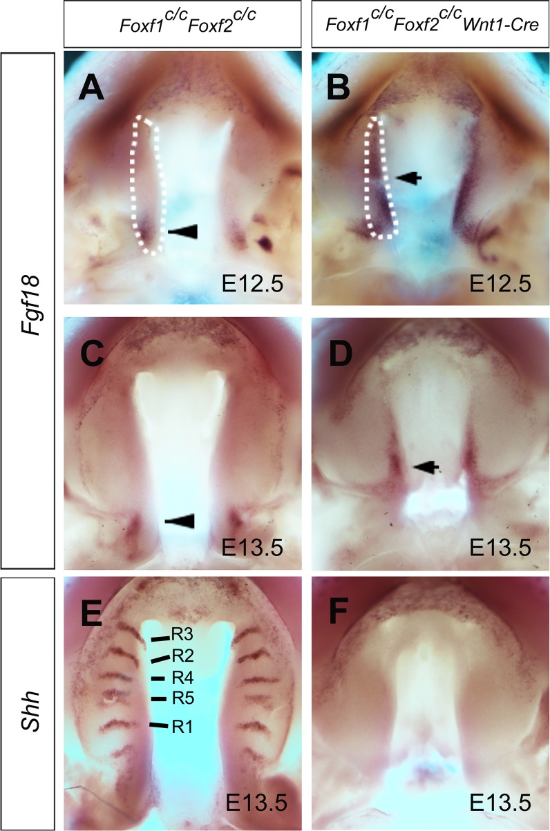
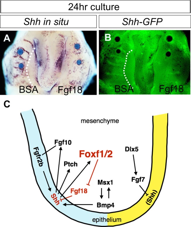
Cleft palate is among the most common birth defects in humans. Previous studies have shown that Shh signaling plays critical roles in palate development and regulates expression of several members of the forkhead-box (Fox) family transcription factors, including Foxf1 and Foxf2, in the facial primordia. Although cleft palate has been reported in mice deficient in Foxf2, whether Foxf2 plays an intrinsic role in and how Foxf2 regulates palate development remain to be elucidated. Using Cre/loxP-mediated tissue-specific gene inactivation in mice, we show that Foxf2 is required in the neural crest-derived palatal mesenchyme for normal palatogenesis. We found that Foxf2 mutant embryos exhibit altered patterns of expression of Shh, Ptch1, and Shox2 in the developing palatal shelves. Through RNA-seq analysis, we identified over 150 genes whose expression was significantly up- or down-regulated in the palatal mesenchyme in Foxf2-/- mutant embryos in comparison with control littermates. Whole mount in situ hybridization analysis revealed that the Foxf2 mutant embryos exhibit strikingly corresponding patterns of ectopic Fgf18 expression in the palatal mesenchyme and concomitant loss of Shh expression in the palatal epithelium in specific subdomains of the palatal shelves that correlate with where Foxf2, but not Foxf1, is expressed during normal palatogenesis. Furthermore, tissue specific inactivation of both Foxf1 and Foxf2 in the early neural crest cells resulted in ectopic activation of Fgf18 expression throughout the palatal mesenchyme and dramatic loss of Shh expression throughout the palatal epithelium. Addition of exogenous Fgf18 protein to cultured palatal explants inhibited Shh expression in the palatal epithelium. Together, these data reveal a novel Shh-Foxf-Fgf18-Shh circuit in the palate development molecular network, in which Foxf1 and Foxf2 regulate palatal shelf growth downstream of Shh signaling, at least in part, by repressing Fgf18 expression in the palatal mesenchyme to ensure maintenance of Shh expression in the palatal epithelium.
Conflict of interest statement
The authors have declared that no competing interests exist.
Figures
Similar articles
-
Genome-wide Identification of Foxf2 Target Genes in Palate Development.J Dent Res. 2020 Apr;99(4):463-471. doi: 10.1177/0022034520904018. Epub 2020 Feb 10. J Dent Res. 2020. PMID: 32040930 Free PMC article.
-
Tissue-specific analysis of Fgf18 gene function in palate development.Dev Dyn. 2021 Apr;250(4):562-573. doi: 10.1002/dvdy.259. Epub 2020 Oct 21. Dev Dyn. 2021. PMID: 33034111 Free PMC article.
-
Altered FGF Signaling Pathways Impair Cell Proliferation and Elevation of Palate Shelves.PLoS One. 2015 Sep 2;10(9):e0136951. doi: 10.1371/journal.pone.0136951. eCollection 2015. PLoS One. 2015. PMID: 26332583 Free PMC article.
-
The Function and Regulatory Network of Pax9 Gene in Palate Development.J Dent Res. 2019 Mar;98(3):277-287. doi: 10.1177/0022034518811861. Epub 2018 Dec 24. J Dent Res. 2019. PMID: 30583699 Review.
-
Gene Regulatory Networks and Signaling Pathways in Palatogenesis and Cleft Palate: A Comprehensive Review.Cells. 2023 Jul 27;12(15):1954. doi: 10.3390/cells12151954. Cells. 2023. PMID: 37566033 Free PMC article. Review.
Cited by
-
Retinoic acid effects on in vitro palatal fusion and WNT signaling.Eur J Oral Sci. 2022 Dec;130(6):e12899. doi: 10.1111/eos.12899. Epub 2022 Oct 27. Eur J Oral Sci. 2022. PMID: 36303276 Free PMC article.
-
Sonic Hedgehog Signaling Is Required for Cyp26 Expression during Embryonic Development.Int J Mol Sci. 2019 May 8;20(9):2275. doi: 10.3390/ijms20092275. Int J Mol Sci. 2019. PMID: 31072004 Free PMC article.
-
Mouse models in palate development and orofacial cleft research: Understanding the crucial role and regulation of epithelial integrity in facial and palate morphogenesis.Curr Top Dev Biol. 2022;148:13-50. doi: 10.1016/bs.ctdb.2021.12.003. Epub 2022 Feb 28. Curr Top Dev Biol. 2022. PMID: 35461563 Free PMC article.
-
Orofacial Cleft and Mandibular Prognathism-Human Genetics and Animal Models.Int J Mol Sci. 2022 Jan 16;23(2):953. doi: 10.3390/ijms23020953. Int J Mol Sci. 2022. PMID: 35055138 Free PMC article. Review.
-
FOXF2 differentially regulates expression of metabolic genes in non-cancerous and cancerous breast epithelial cells.Trends Diabetes Metab. 2018;1(1):10.15761/TDM.1000103. doi: 10.15761/TDM.1000103. Epub 2018 Jul 6. Trends Diabetes Metab. 2018. PMID: 30294731 Free PMC article.
References
-
- Ferguson MW. Palate development. Development. 1988;103 Suppl:41–60. - PubMed
-
- Gritli-Linde A. Molecular control of secondary palate development. Developmental biology. 2007;301(2):309–326. - PubMed
-
- Chai Y, Maxson RE Jr. Recent advances in craniofacial morphogenesis. Developmental dynamics: an official publication of the American Association of Anatomists. 2006;235(9):2353–2375. - PubMed
-
- Rice R, Connor E, Rice DP. Expression patterns of Hedgehog signalling pathway members during mouse palate development. Gene expression patterns: GEP. 2006;6(2):206–212. - PubMed
Publication types
MeSH terms
Substances
Grants and funding
LinkOut - more resources
Full Text Sources
Other Literature Sources
Molecular Biology Databases
