

Population of 224 realistic human subject-based computational breast phantoms
- PMID: 26745896
- PMCID: PMC4684566
- DOI: 10.1118/1.4937597
Population of 224 realistic human subject-based computational breast phantoms
Abstract
Purpose: To create a database of highly realistic and anatomically variable 3D virtual breast phantoms based on dedicated breast computed tomography (bCT) data.
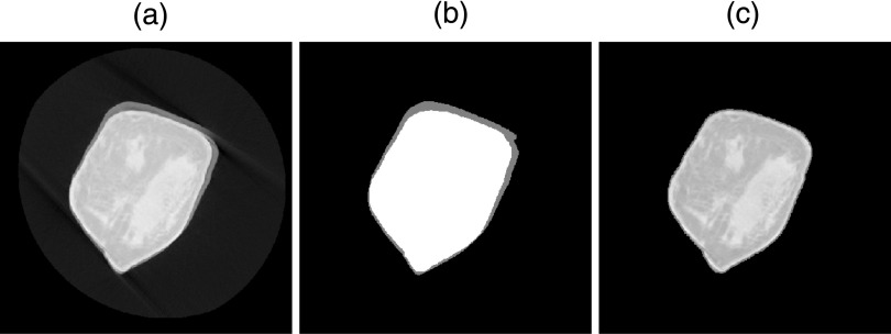
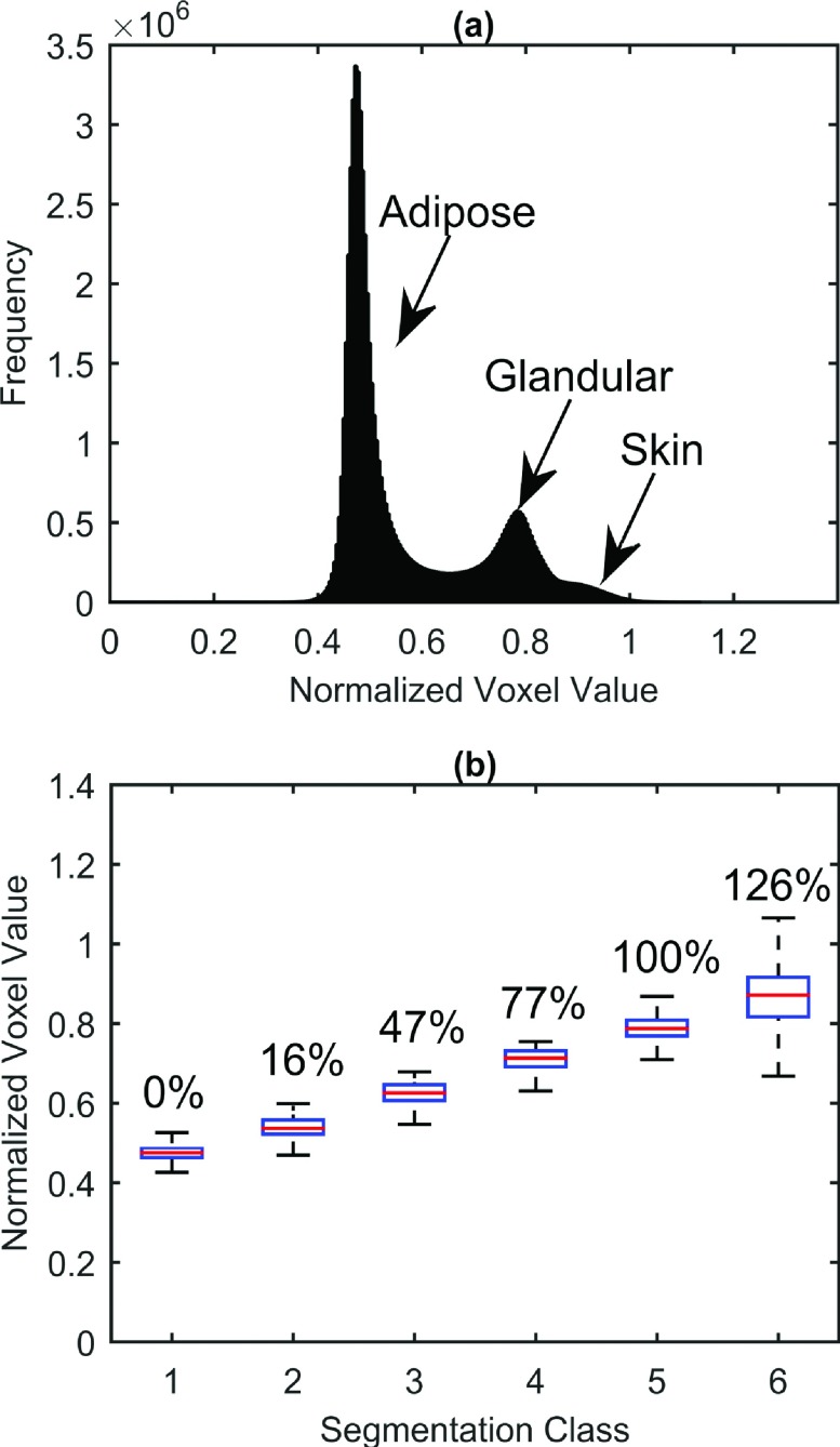
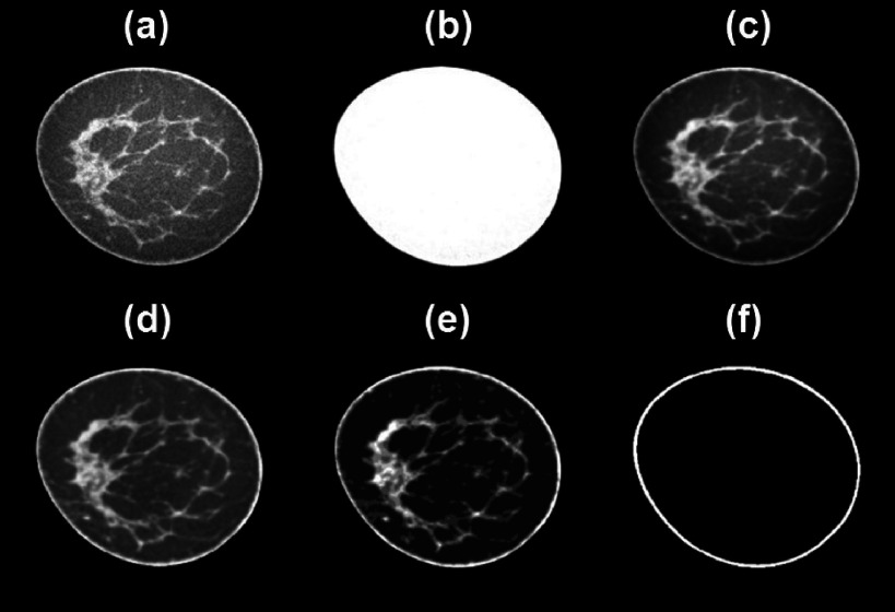
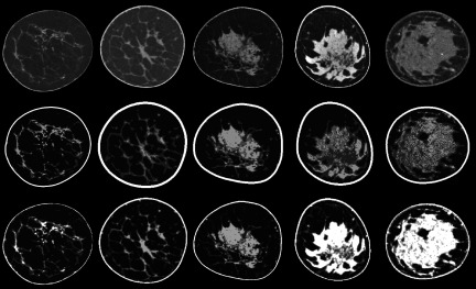
Methods: A tissue classification and segmentation algorithm was used to create realistic and detailed 3D computational breast phantoms based on 230 + dedicated bCT datasets from normal human subjects. The breast volume was identified using a coarse three-class fuzzy C-means segmentation algorithm which accounted for and removed motion blur at the breast periphery. Noise in the bCT data was reduced through application of a postreconstruction 3D bilateral filter. A 3D adipose nonuniformity (bias field) correction was then applied followed by glandular segmentation using a 3D bias-corrected fuzzy C-means algorithm. Multiple tissue classes were defined including skin, adipose, and several fractional glandular densities. Following segmentation, a skin mask was produced which preserved the interdigitated skin, adipose, and glandular boundaries of the skin interior. Finally, surface modeling was used to produce digital phantoms with methods complementary to the XCAT suite of digital human phantoms.
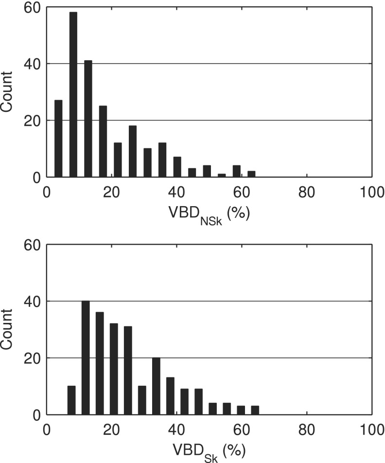
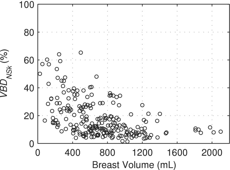
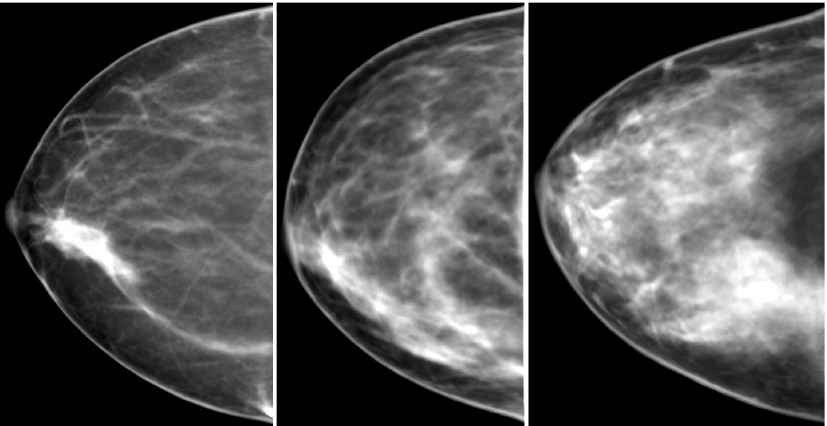
Results: After rejecting some datasets due to artifacts, 224 virtual breast phantoms were created which emulate the complex breast parenchyma of actual human subjects. The volume breast density (with skin) ranged from 5.5% to 66.3% with a mean value of 25.3% ± 13.2%. Breast volumes ranged from 25.0 to 2099.6 ml with a mean value of 716.3 ± 386.5 ml. Three breast phantoms were selected for imaging with digital compression (using finite element modeling) and simple ray-tracing, and the results show promise in their potential to produce realistic simulated mammograms.
Conclusions: This work provides a new population of 224 breast phantoms based on in vivo bCT data for imaging research. Compared to previous studies based on only a few prototype cases, this dataset provides a rich source of new cases spanning a wide range of breast types, volumes, densities, and parenchymal patterns.
Figures
References
-
- Kerlikowske K., Carney P. A., Geller B., Mandelson M. T., Taplin S. H., Malvin K., Ernster V., Urban N., Cutter G., Rosenberg R., and Ballard-Barbash R., “Performance of screening mammography among women with and without a first-degree relative with breast cancer,” Ann. Intern. Med. 133, 855–863 (2000). 10.7326/0003-4819-133-11-200012050-00009 - DOI - PubMed
Publication types
MeSH terms
Grants and funding
LinkOut - more resources
Full Text Sources
Other Literature Sources
Medical
