. 2016 Feb 15;196(4):1666-77.
doi: 10.4049/jimmunol.1501870. Epub 2016 Jan 8.

IRF4 and IRF8 Act in CD11c+ Cells To Regulate Terminal Differentiation of Lung Tissue Dendritic Cells

Affiliations

IRF4 and IRF8 Act in CD11c+ Cells To Regulate Terminal Differentiation of Lung Tissue Dendritic Cells

Sandra Bajaña et al. J Immunol. .

Abstract

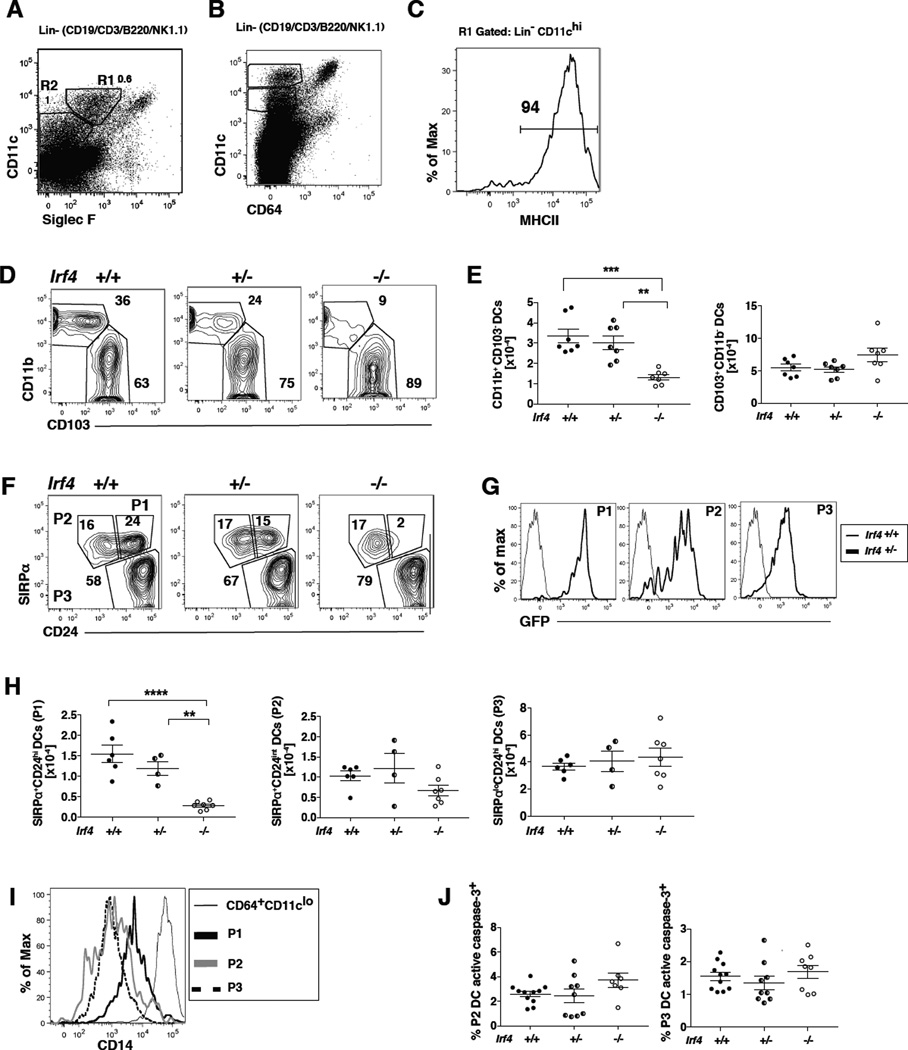
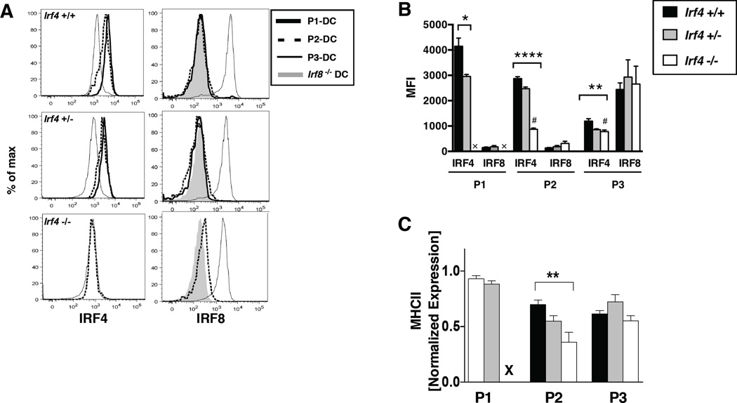
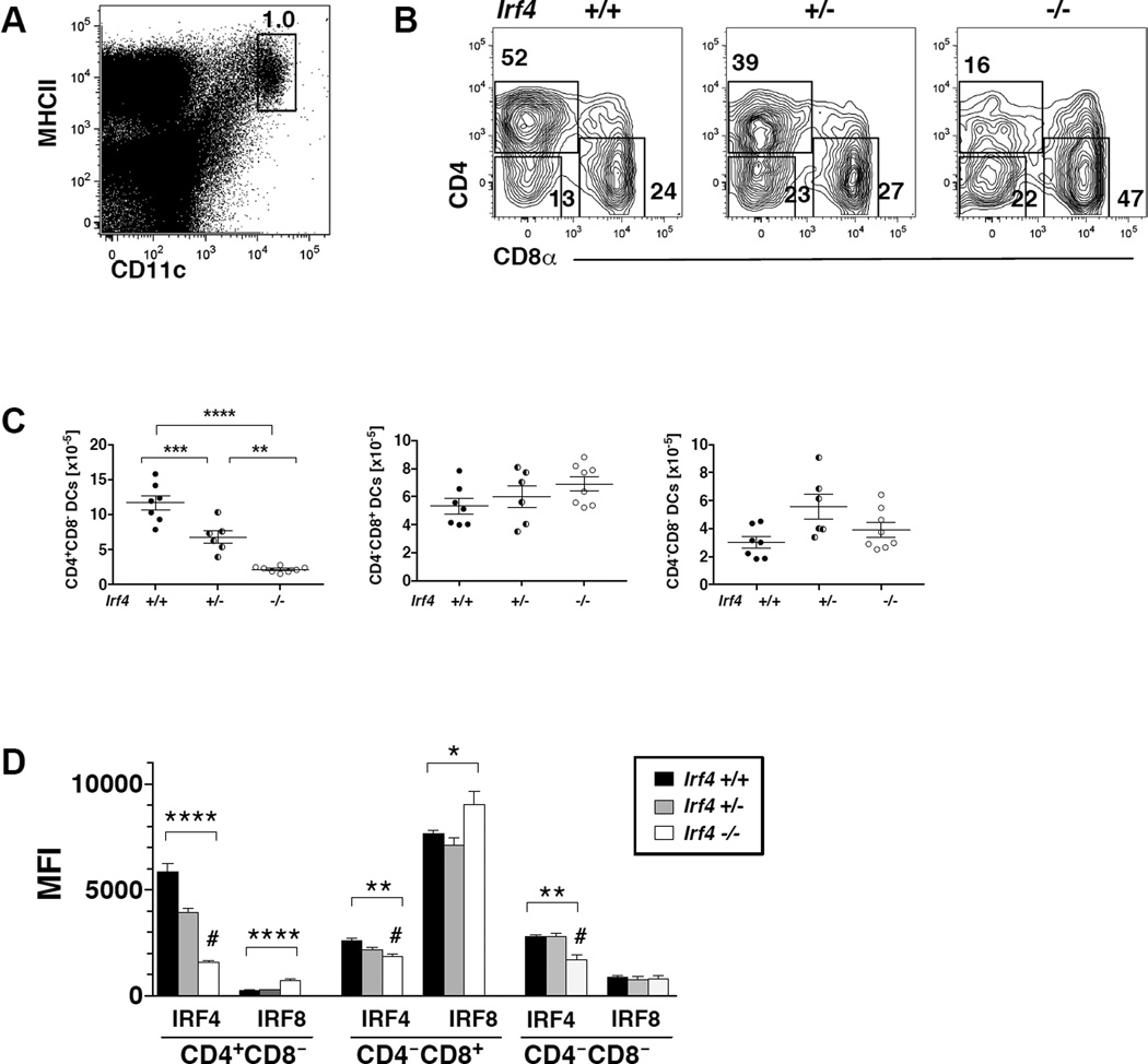
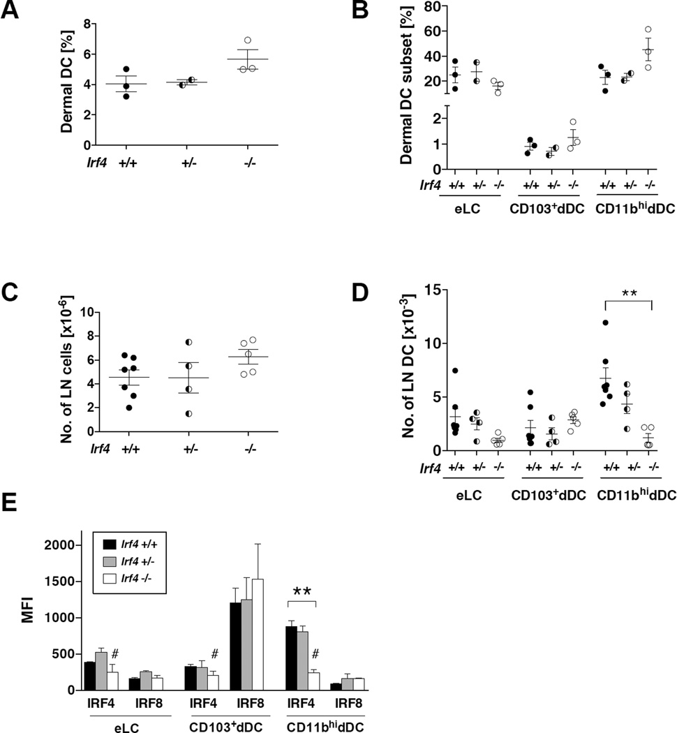
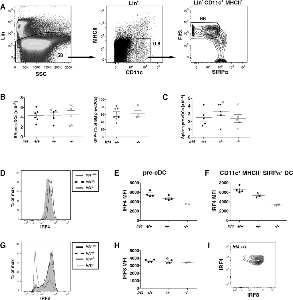
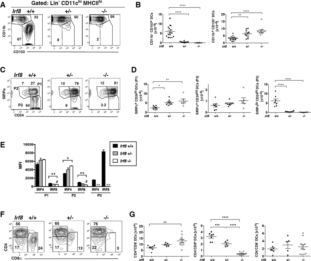
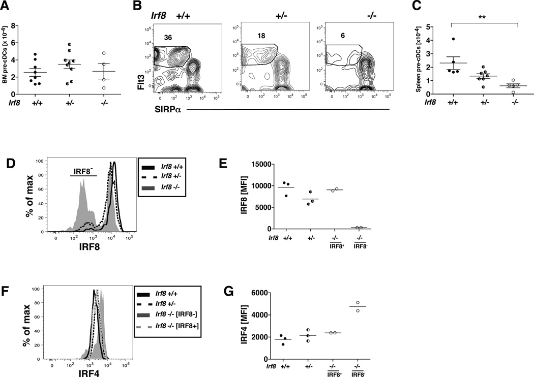
Dendritic cells (DCs) initiate immune responses in barrier tissues including lung and skin. Conventional DC (cDC) subsets, CD11b(-) (cDC1s) or CD11b(+) (cDC2s), arise via distinct networks of transcription factors involving IFN regulatory factor 4 (IRF4) and IRF8, and are specialized for unique functional responses. Using mice in which a conditional Irf4 or Irf8 allele is deleted in CD11c(+) cells, we determined whether IRF4 or IRF8 deficiency beginning in CD11c(+) cDC precursors (pre-cDCs) changed the homeostasis of mature DCs or pre-DCs in the lung, dermis, and spleen. CD11c-cre-Irf4(-/-) mice selectively lacked a lung-resident CD11c(hi)CD11b(+)SIRPα(+)CD24(+) DC subset, but not other lung CD11b(+) DCs or alveolar macrophages. Numbers of CD11b(+)CD4(+) splenic DCs, but not CD11b(+) dermal DCs, were reduced, indicating cDC2s in the lung and dermis develop via different pathways. Irf4 deficiency did not alter numbers of cDC1s. CD11c-cre-Irf8(-/-) mice lacked lung-resident CD103(+) DCs and splenic CD8α(+) DCs, yet harbored increased IRF4-dependent DCs. This correlated with a reduced number of Irf8(-/-) pre-cDCs, which contained elevated IRF4, suggesting that Irf8 deficiency diverts pre-cDC fate. Analyses of Irf4 and Irf8 haploinsufficient mice showed that, although one Irf4 allele was sufficient for lung cDC2 development, two functional Irf8 alleles were required for differentiation of lung cDC1s. Thus, IRF8 and IRF4 act in pre-cDCs to direct the terminal differentiation of cDC1 and cDC2 subsets in the lung and spleen. These data suggest that variation in IRF4 or IRF8 levels resulting from genetic polymorphisms or environmental cues will govern tissue DC numbers and, therefore, regulate the magnitude of DC functional responses.


Figures

Fig. 1
Fig. 1. IRF4 expression in CD11c+ cells is required for development of a CD11chiMHCIIhiCD11b+CD24hiSIRPα+ DC subset in the lung
(A–C) Definition of DC populations in CD11c-cre-Irf4 +/+ mice. (A) CD11c+ myeloid cells displaying distinct levels of SiglecF are identified in the Lineage-negative (CD19CD3B220 NK1.1) fraction of lung cells. The R1 gate defines CD11chiSiglecFlo DCs that are (B) CD64 and (C) MHCIIhi. The R2 gate defines CD11cintSiglecF cells, some of which are (B) CD64 and MHCII+ (see Fig. S2). The numbers within these plots from +/+ mice indicate the percentage of cells within each gate. (D) CD11chiMHCIIhi cells in gate R1 are divided into CD11b+ and CD103+ subsets; a comparison of +/+, +/− and −/− mice is shown. (E) The total numbers of CD11b+ and CD103+ DC subsets in multiple +/+, +/− and −/− mice are compiled; shown are values for individual mice, n=7 per genotype. (F) CD11chiMHCIIhi cells in gate R1 are divided into P1, P2 and P3 subsets based on SIRPα and CD24; a comparison of +/+, +/− and −/− mice is shown. (G) The CD11chiMHCIIhi DC subsets SIRPα+CD24hi (P1), SIRPα+CD24int (P2) and SIRPαloCD24hi (P3) are GFP+ in +/− mice. (H) The total numbers of P1, P2 and P3 DC subsets in multiple +/+, +/− and −/− mice are compiled, n=4–7 per genotype. (I) DCs within the CD11chi P1, P2 and P3 subsets display lower levels of CD14 than the CD11clo CD64hi macrophages. (J) Shown is the percentage of DCs in P2 and P3 subsets that contain activated caspase-3 in +/+, +/− and −/− mice, n=8–11 per genotype. The significance of the data in panels E, H and J was evaluated using a one-way ANOVA with a Tukey multiple comparisons test; **p<0.01, ***p<0.001, ****p<0.0001.
Fig. 2
Fig. 2. cDC subsets in CD11c-cre-Irf4 mice differ in expression of IRF4 and IRF8 while heterozygotes show reduced levels of protein
(A) In CD11c-cre-Irf4 mice, lung cDCs were subdivided into P1, P2 and P3 subsets as in Fig. 1F, and intracellular IRF4 and IRF8 levels were determined using flow cytometry. For each subset in +/+, +/− and −/− mice, the binding of anti-IRF4 (left panels) and anti-IRF8 (right panels) is shown. The CD11c-cre-Irf4 −/− and CD11c-cre-Irf8 −/− mice were used to determine the nonspecific level of anti-IRF4 and anti-IRF8 Ab binding, respectively, to DCs. (B) The mean fluorescence intensity (MFI) of anti-IRF4 and anti-IRF8 binding is shown for each cDC subset (P1, P2, P3) in CD11c-cre-Irf4 +/+, +/− and −/− mice. The # indicates the nonspecific binding of the anti-IRF4 Ab on Irf4 −/− cells. X indicates that the P1 subset is absent in the −/− mouse. (C) The relative MHCII expression (normalized MFI) on the P1, P2 and P3 subsets present in CD11c-cre-Irf4 +/+, +/− and −/− mice is shown. X indicates that the P1 subset is absent in the −/− mouse. For panels B and C, the significance of the data was evaluated using an unpaired t test (P1) or a one-way ANOVA (P2, P3); *p<0.05, **p<0.01, ****p<0.0001, n=4 per genotype.
Fig. 3
Fig. 3. IRF4 expression in CD11c+ cells is required for development of splenic CD11b+CD4+DCs
(A) Definition of CD11chiMHCIIhi cDCs in the spleen of CD11c-cre-Irf4 +/+ mice. (B) CD11chiMHCIIhi cells are divided into CD4+CD8, CD4CD8+ and CD4CD8 DC subsets; a comparison of +/+, +/− and −/− mice is shown. (C) The total numbers of CD4+CD8, CD4CD8+ and CD4CD8 DC subsets in multiple +/+, +/− and −/− mice are compiled; shown are values for individual mice, n=6–8 per genotype. The significance of the data was evaluated using a one-way ANOVA with a Tukey multiple comparisons test; **p<0.01, ***p<0.001, ****p<0.0001. (D) The mean fluorescence intensity (MFI) of anti-IRF4 and anti-IRF8 binding is shown for the CD4+CD8, CD4CD8+ and CD4CD8 DC subsets in +/+, +/− and −/− mice. The # indicates the background binding of the anti-IRF4 Ab on Irf4−/− cells. The binding of anti-IRF8 to Irf8−/− splenic DCs was a MFI of 195. The significance of the data was evaluated using a one-way ANOVA; *p<0.05, **p<0.01, ****p<0.0001, n=4 per genotype.
Fig. 4
Fig. 4. Irf4 deficiency in CD11c+ cells does not affect skin DC development but reduces migration of CD11b+ dermal DCs to local draining lymph nodes
(A–B) The percentages of total dermal DCs and distinct DC subsets (eLC, CD103+, CD11bhi) in the skin of multiple CD11c-cre-Irf4 +/+, +/− and −/− mice, n=2–3. (C–D) Numbers of LN cells and migratory DC subsets in CD11c-cre-Irf4 +/+, +/− and −/− mice, n=4–7. (E) The MFI of anti-IRF4 and anti-IRF8 binding is shown for the eLC, CD103+ and CD11bhi DC subsets in +/+, +/− and −/− mice. The # indicates the background binding of the anti-IRF4 Ab on Irf4−/− cells. The binding of anti-IRF8 to Irf8−/− CD11bhi DCs was a MFI of 102. The significance of the data was evaluated with a one-way ANOVA with a Tukey’s multiple comparisons test, **p<0.01.
Fig. 5
Fig. 5. IRF4 deficiency in CD11c+ cells does not alter numbers or IRF8 expression of pre-cDCs in bone marrow or spleen
(A) Definition of pre-cDCs in the bone marrow of CD11c-cre-Irf4 +/+ mice as lineage-negative (Lin, CD19CD3NK1.1Ter119B220) CD11c+MHCIISIRPαloFlt3+. (B–C) The numbers of total pre-cDCs and GFP+ pre-cDCs in the bone marrow and spleen in multiple +/+, +/− and −/− mice are compiled, n=4–6 per genotype. (D) IRF4 protein levels were determined in +/+, +/− and −/− BM pre-cDCs by intracellular staining, and (E) the anti-IRF4 MFI values were compiled from multiple mice. (F) IRF4 protein levels were determined in CD11c+MHCII+SIRPα+ cDCs in BM of +/+, +/− and −/− mice by intracellular staining, and the anti-IRF4 MFI values were compiled from multiple mice. (G) IRF8 protein levels were determined in +/+, +/− and −/− pre-cDCs by intracellular staining, and (H) the anti-IRF8 MFI values were compiled from multiple mice. Binding of the anti-IRF8 mAb to pre-cDCs of Irf8-deficient mice was used to show background levels of mAb binding, and this revealed that not all pre-cDCs in the Irf8-deficient mice have deleted Irf8 (see Fig. 7). (I) Binding of anti-IRF4 and anti-IRF8 mAbs to pre-cDCs in +/+ mice.
Fig. 6
Fig. 6. IRF8 deficiency in CD11c+ cells alters numbers of both IRF8- and IRF4-dependent lung and spleen DCs
(A) CD11chiMHCIIhi DCs (gate R1) in the lungs of CD11c-cre-Irf8 mice were gated as in Fig. 1A and divided into CD11b+ and CD103+ subsets; a comparison of +/+, +/− and −/− mice is shown. (B) The total numbers of CD11b+ and CD103+ DC subsets in multiple +/+, +/− and −/− mice are compiled; shown are values for individual mice, n=10–15 per genotype. (C) Lung CD11chiMHCIIhi cells in gate R1 (see Fig. 1A) are divided into subsets based on SIRPα and CD24; a comparison of +/+, +/− and −/− mice is shown. (D) The total numbers of SIRPα+CD24hi (P1), SIRPα+CD24int (P2) and SIRPαloCD24hi (P3) DC subsets in multiple +/+, +/− and −/− mice are compiled, n=7–8 per genotype. For panels B and D, the significance of the data was evaluated using a one-way ANOVA with a Tukey’s multiple comparisons test; *p<0.05, **p<0.01, ****p<0.0001. (E) The mean fluorescence intensity (MFI) of anti-IRF4 and anti-IRF8 binding is shown for each lung cDC subset (P1, P2, P3) in CD11c-cre-Irf8 +/+, +/− and −/− mice. The # indicates the nonspecific binding of the anti-IRF8 Ab to Irf8 −/− cells. X indicates that the P3 subset is absent in the +/− and −/− mice. The significance of the data was evaluated using a one-way ANOVA (P1, P2); *p<0.05, **p<0.01, n=3 per genotype. (F) Splenic CD11chiMHCIIhi cells (gated as in Fig. 3A) are divided into CD4+ and CD8α+ subsets; a comparison of +/+, +/− and −/− mice is shown. (G) The total numbers of CD4+ and CD8α+ splenic DC subsets in multiple +/+, +/− and −/− mice are compiled; shown are values for individual mice, n=5–11 per genotype.
Fig. 7
Fig. 7. IRF8 deficiency in CD11c+ cells leads to decreased numbers of splenic pre-cDCs expressing elevated levels of IRF4
(A) The numbers of pre-cDCs in the bone marrow of multiple CD11c-cre-Irf8 +/+, +/− and −/− mice are compiled, n=4–9 per genotype. (B) Shown is the gating of SIRPαFlt3+ pre-cDCs within the lineage-negative (CD19CD3NK1.1Ter119B220) CD11c+MHCII fraction of splenocytes in CD11c-cre-Irf8 +/+, +/− and −/− mice. (C) The numbers of pre-cDCs in the spleens of multiple CD11c-cre-Irf8 +/+, +/− and −/− mice are compiled, n=5–7 per genotype. (D) IRF8 protein levels were determined in +/+, +/− and −/− pre-cDCs by intracellular staining. Binding of the anti-IRF8 mAb to pre-cDCs of Irf8-deficient mice revealed that not all pre-cDCs in the Irf8-deficient mice have deleted Irf8. (E) The anti-IRF8 MFI values were compiled from multiple mice (n=2–3). In −/− mice, the IRF8 MFI is reported separately for the IRF8+ and IRF8 populations. (F) IRF4 protein levels were determined in +/+, +/− and −/− pre-cDCs by intracellular staining, and (G) the anti-IRF4 MFI values were compiled from multiple mice (n=2–3). In −/− mice, the IRF4 MFI is reported separately for the IRF8+ and IRF8 populations.

References

    1. Braciale TJ, Sun J, Kim TS. Regulating the adaptive immune response to respiratory virus infection. Nat Rev Immunol. 2012;12:295–305. - PMC - PubMed
    1. Lambrecht BN, Hammad H. Lung dendritic cells in respiratory viral infection and asthma: from protection to immunopathology. Annu Rev Immunol. 2012;30:243–270. - PubMed
    1. Satpathy AT, Wu X, Albring JC, Murphy KM. Re(de)fining the dendritic cell lineage. Nat Immunol. 2012;13:1145–1154. - PMC - PubMed
    1. Belz GT, Nutt SL. Transcriptional programming of the dendritic cell network. Nat Rev Immunol. 2012;12:101–113. - PubMed
    1. Miller JC, Brown BD, Shay T, Gautier EL, Jojic V, Cohain A, Pandey G, Leboeuf M, Elpek KG, Helft J, Hashimoto D, Chow A, Price J, Greter M, Bogunovic M, Bellemare-Pelletier A, Frenette PS, Randolph GJ, Turley SJ, Merad M. Deciphering the transcriptional network of the dendritic cell lineage. Nat Immunol. 2012;13:888–899. - PMC - PubMed

Publication types

Substances