. 2016 Jan 8:17:5.
doi: 10.1186/s12882-015-0216-0.

Cells of renin lineage express hypoxia inducible factor 2α following experimental ureteral obstruction

Affiliations

Cells of renin lineage express hypoxia inducible factor 2α following experimental ureteral obstruction

Ania Stefanska et al. BMC Nephrol. .

Abstract

Background: Recent studies indicate that mural cells of the preglomerular vessels, known as cells of renin lineage (CoRL), contribute to repair and regeneration of injured kidney glomeruli. However, their potential roles in tubulointerstitial disease are less understood. The aim of this study was to better understand CoRL number and distribution following UUO so that future mechanistic studies could be undertaken.

Methods: We mapped the fate of CoRL in adult Ren1cCreER x Rs-tdTomato-R reporter mice that underwent UUO. Kidney biopsies from sham and UUO-subjected mice on days 3, 7, and 14 were evaluated by immunohistochemistry.

Results: In sham animals, CoRL were restricted to juxtaglomerular location. At day 7 following UUO, CoRL increased two-fold, were perivascular in location, and co-expressed pericyte markers (PDGFßR, NG2), but did not express renin. At day 14 post UUO, labeled CoRL detached from vessels and were present in the interstitium, in areas of fibrosis, where they now expressed the myofibroblast marker alpha-smooth muscle actin. The increase in CoRL was likely due to proliferation as marked by BrdU labeling, and migration from the cortex. Following UUO starting from day 3, active hypoxia inducible factor-2α was detected in nuclei in labeled CoRL, in the cortex, but not those cells found in medulla.

Conclusions: We have demonstrated that arteriolar CoRL are potential kidney progenitors that may contribute to the initial vascular regeneration. However, in chronic kidney injury (≥14 days post UUO), perivascular CoRL transition to myofibroblast-like cells.


Figures
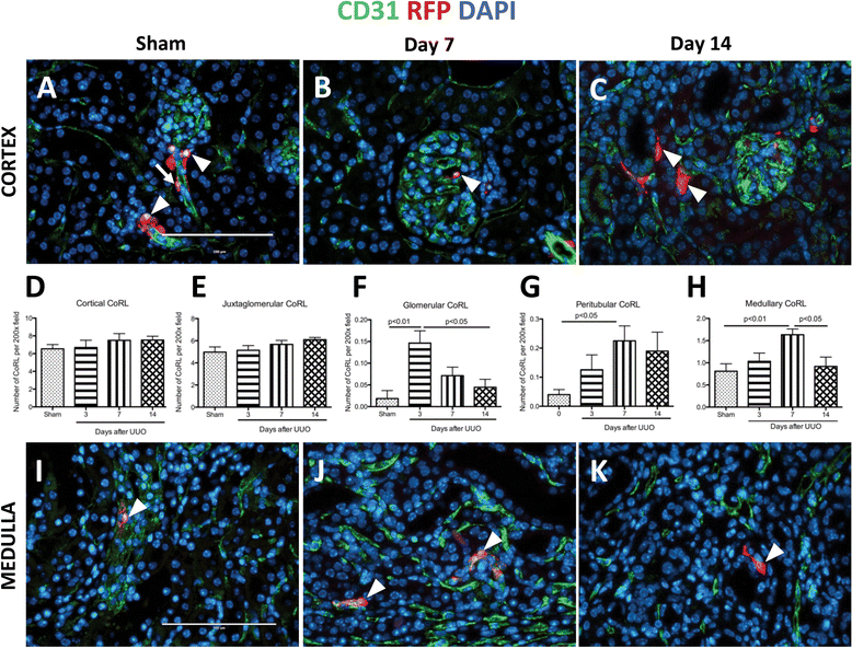
Fig. 1
Fig. 1
The number of cells of renin lineage (CoRL) increases beyond the JG location following UUO. CoRL were identified by RFP reporter staining (red), endothelial cells were labeled with CD31 staining (green). Nuclei are labeled with DAPI (blue). (ag) CoRL in Cortex. (a) Representative images show that in sham-operated mice, labeled CoRL (arrowheads) are predominantly localized to the juxta-glomerulus, with fewer detected along afferent arterioles (arrow). (b) At d7 post UUO, occasional labeled CoRL were detected in glomerular capillary loops. (c) Labeled CoRL were rarely detected in peritubular locations at day 14. Quantification: (d) Total cortical CoRL number was not statistically significant following UUO, (e) the same was found when CoRL were quantified exclusively in JG compartment. However, (f) there was a significant increase of intraglomerular CoRL at d3, and (g) peritubular CoRL at d7 and d14. (hk) CoRL in Medulla. (h) Quantification - total CoRL number in the medulla was significantly increased on day 7 following UUO. (i) In the medulla of the sham kidney, CoRL were restricted to vasa recta. (j) Following UUO there was an increase of CoRL in vasa recta on day 7 (arrowheads). (k) 14 days after UUO, a subset of CoRL localized away from any vessels, being present in the interstitium
Fig. 2
Fig. 2
Proliferative and migratory phenotype of CoRL in UUO. (ac) To detect proliferation in CoRL, double-staining was performed for Red fluorescent protein (red) to identify CoRL, and BrdU (green) to examine cell proliferation. Nuclei are labeled with DAPI (blue). Medulla: (a) CoRL did not co-express BrdU in sham kidneys. ( b) At day 7 post UUO, a subset of CoRL co-expressed BrdU (yellow color; arrow shows example). The inset demonstrates this at higher magnification. As expected, other peritubular cells stained for BrdU (open arrowheads). (c) Some dual CoRL+/BrdU+ cells displayed elongated shape with long cellular processes suggesting cell motility when viewed at high power (inset). (d) To better define the subset of labeled CoRL that appeared to be migrating based on their shape (identified by RFP staining, arrowheads), double staining was performed for the endothelial cell marker CD31 (green, arrows). (d’) On occasion, CoRL were detected within vessels in the medulla
Fig. 3
Fig. 3
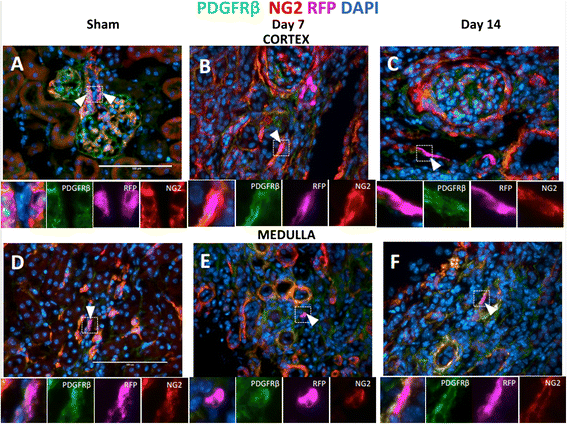
Interstitial CoRL co-express pericytes markers. To label pericytes, antibody staining against PDGFRß (green) and NG2 (red) was performed, CoRL were identified by RFP staining, nuclei are labeled with DAPI. (a c) Cortex: (a) Representative images show that in sham-operated mice, CoRL co-express pericyte markers in JG (arrowhead). Insets show high power images of juxtaglomerular CoRL. In UUO, there was an accumulation of interstitial pericytes (orange color). Interstitial CoRL were found in peritubular areas co-expressing pericyte markers at (b) 7 day post UUO, and (c) 14 days post UUO. Insets show high power images of CoRL. (df) Medulla: (d) Sham medulla shows CoRL co-expressing pericyte markers in vasa recta (arrowheads). Insets show high power images. Pericyte marker staining increases in UUO (orange color). Interstitial CoRL were co-labeled with pericyte markers at (e) day 7 post UUO, (f) day 14 post UUO. Insets show high power images of interstitial CoRL
Fig. 4
Fig. 4
αSMA is expressed by interstitial CoRL in fibrosis. Red fluorescent protein (red) staining identified CoRL, αSMA (green) staining was used to mark vascular smooth muscle cells and myofibroblasts, CD31 staining denotes endothelial cells (magenta). Nuclei are labeled with DAPI (blue). (a) In the sham cortex, CoRL were found in JG compartment (arrowheads, insets). (b) At d7 post UUO, CoRL were localized to perivascular area (arrowhead, insets), however, some CoRL were no longer supporting microvessels (arrow). (c) At 14 days post UUO, majority of CoRL (arrowhead) were found outside vascular bed and expressing αSMA suggesting the myofibroblastic conversion. Insets show high power images of merged and single stained panels (d) In the sham medulla, CoRL were found in vasa recta (arrowhead) and co-expressing αSMA. (e) 7 days following UUO, CoRL were still found surrounding endothelial cells (arrowhead, insets). (f) 14 days post UUO, CoRL and localized to avascular areas and expressing αSMA (arrowhead). Insets show high power images of merged and single stained panels
Fig. 5
Fig. 5
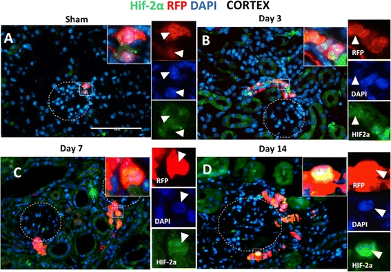
Nuclear translocation of HIF-2α in JG and afferent arteriole in UUO. Red fluorescent protein (red) staining identified CoRL, HIF-2α (green) staining was used to detect hypoxia-activated cells. Nuclei are labeled with DAPI (blue). In the sham kidney cortex, there was (a) a cytoplasmic HIF-2α staining in JG cells (inset shows single panel colors, arrowheads indicates the same cell). In UUO kidney, there was an accumulation of HIF-2α staining in the nuclei in afferent arterioles and JG (insets show single panel colors, arrowheads indicates the same cell) on (b) day 3, (c) day 7, (d) day 14 following kidney injury

References

    1. Chevalier R, Forbes M, Thornhill B. Ureteral obstruction as a model of renal interstitial fibrosis and obstructive nephropathy. Kidney Int. 2009;75:1145–52. doi: 10.1038/ki.2009.86. - DOI - PubMed
    1. Ucero AC, Benito-Martin A, Izquierdo MC, Sanchez-Niño MD, Sanz AB, Ramos AM, et al. Unilateral ureteral obstruction: beyond obstruction. Int Urol Nephrol. 2014;46:765–76. doi: 10.1007/s11255-013-0520-1. - DOI - PubMed
    1. Moody TE, Vaughn ED, Gillenwater JY. Relationship between renal blood flow and ureteral pressure during 18 hours of total unilateral uretheral occlusion. Implications for changing sites of increased renal resistance. Invest Urol. 1975;13:246–51. - PubMed
    1. Ucero AC, Gonçalves S, Benito-Martin A, Santamaría B, Ramos AM, Berzal S, et al. Obstructive renal injury: from fluid mechanics to molecular cell biology. Open access J Urol. 2010;2:41–55. - PMC - PubMed
    1. Franco M, Roswall P, Cortez E, Hanahan D, Pietras K. Pericytes promote endothelial cell survival through induction of autocrine VEGF-A signaling and Bcl-w expression. Blood. 2011;118:2906–17. doi: 10.1182/blood-2011-01-331694. - DOI - PMC - PubMed

Publication types

MeSH terms

Substances