. 2017 Jan;37(1):291-306.
doi: 10.1177/0271678X15624933. Epub 2016 Jan 8.

Neuronal HIF-1α and HIF-2α deficiency improves neuronal survival and sensorimotor function in the early acute phase after ischemic stroke

Affiliations

Neuronal HIF-1α and HIF-2α deficiency improves neuronal survival and sensorimotor function in the early acute phase after ischemic stroke

Philipp Barteczek et al. J Cereb Blood Flow Metab. 2017 Jan.

Abstract

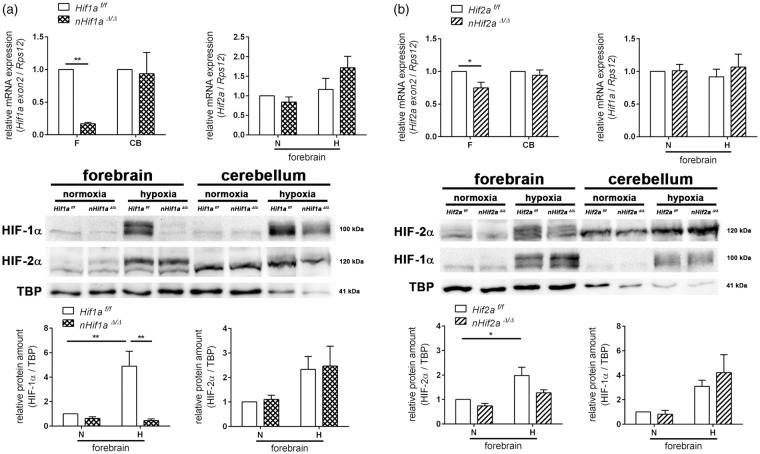
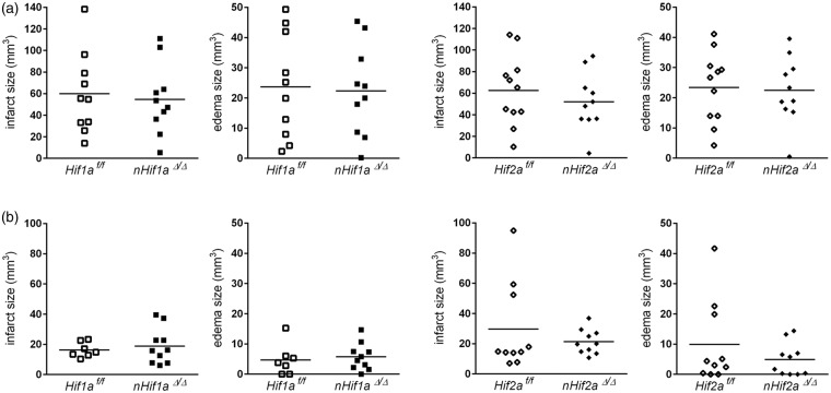
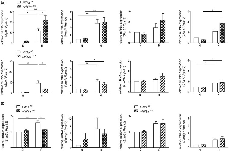
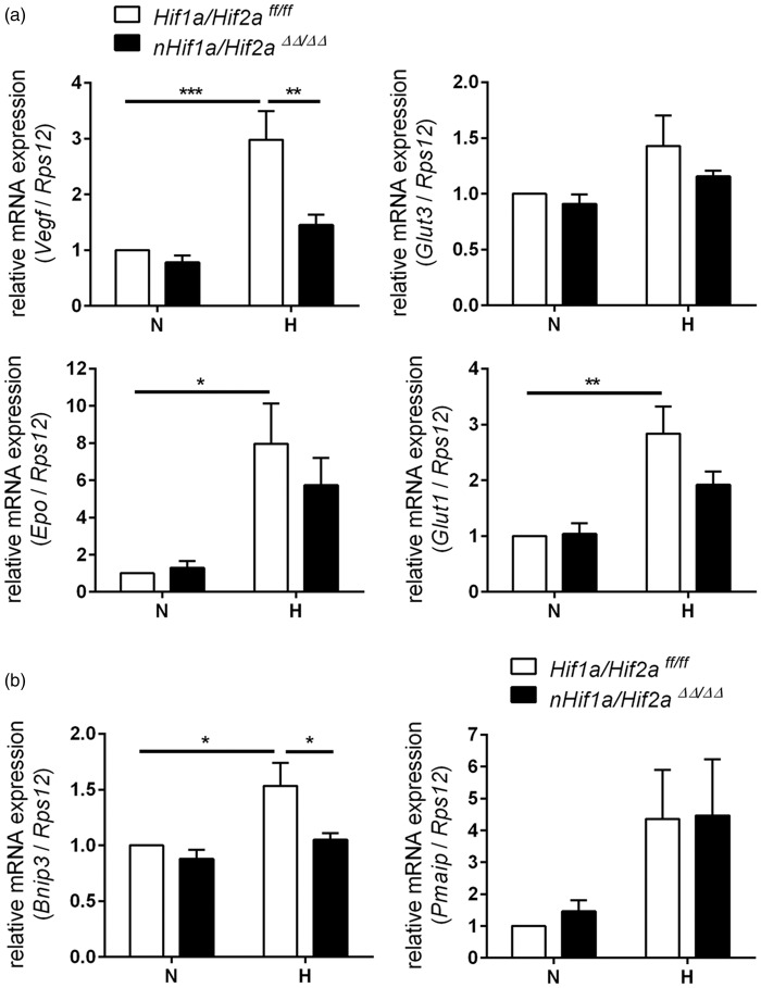
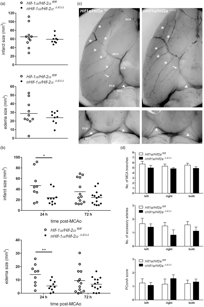
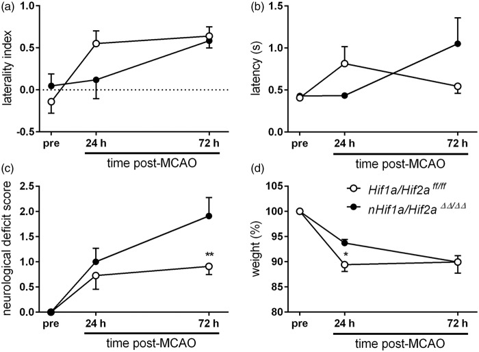
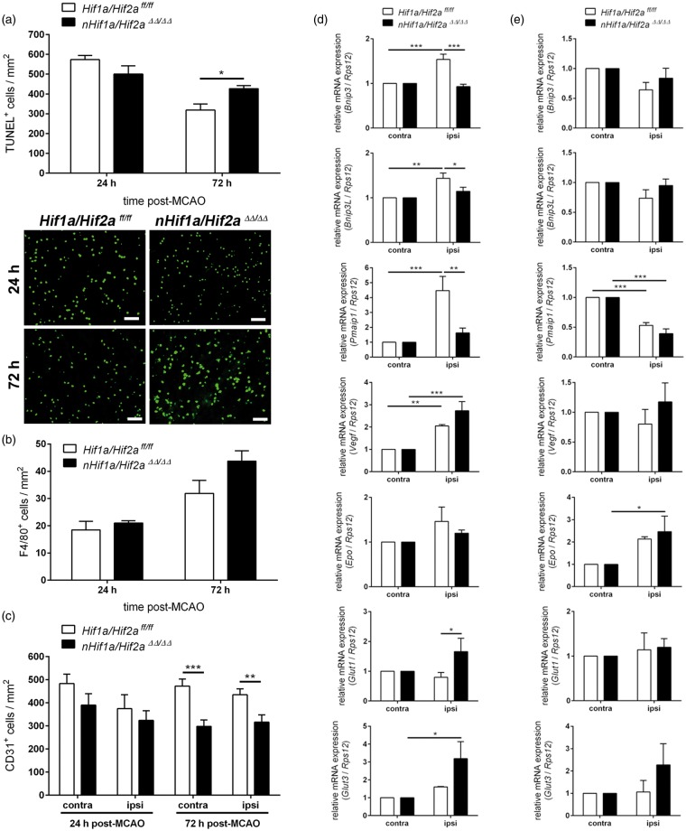
Hypoxia-inducible factors mediate adaptive responses to ischemia, among others, by induction of anti- and pro-survival genes. Thus, the impact of HIF on neuronal survival upon stroke is controversial. Therefore, neuron-specific knockout mice deficient for Hif1a and Hif2a were exposed to inspiratory hypoxia or ischemia-reperfusion injury. Both Hif1a- and Hif2a-deficient mice showed no altered infarct and edema size, suggesting that both HIF-α subunits might compensate for each other. Accordingly, hypoxic HIF-target gene regulation was marginally affected with exception of anti-survival Bnip3 and pro-survival erythropoietin. In the early acute stage upon stroke, Hif1a/Hif2a double knockout mice exhibited significantly reduced expression of the anti-survival Bnip3, Bnip3L, and Pmaip1 Accordingly, global cell death and edema were significantly reduced upon 24 h but not 72 h reperfusion. Behavioral assessment indicated that Hif1a/Hif2a-deficient mice initially performed better, but became significantly more impaired after 72 h accompanied by increased apoptosis and reduced angiogenesis. Our findings suggest that in neurons HIF-1 and HIF-2 have redundant functions for cellular survival under ischemic conditions. By contrast, lack of anti-survival factors in Hif1a/Hif2a-deficient mice might protect from early acute neuronal cell death and neurological impairment, indicating a benefit of HIF-pathway inhibition in neurons in the very acute phase after ischemic stroke.

Keywords: Erythropoietin; hypoxia-inducible factor; ischemia; stroke; vascular endothelial growth factor.


Figures

Figure 1.
Figure 1.
Characterization of neuron-specific Hif1a and Hif2a knockout. Hif1af/f and nHif1aΔ/Δ (a) or Hif2af/f and nHif2aΔ/Δ (b) mice were exposed to normobaric hypoxia (6% oxygen) for 6 h or were kept at normoxic conditions. Nuclear proteins and RNA were prepared from forebrain and cerebellum. Gene expression was determined by real-time polymerase chain reaction (PCR) (n = 5, per group) and Western blotting (n = 3–4, per group). Values are normalized to Rps12 (mRNA) or TBP (protein) and expressed as fold change to normoxic Hif1af/f and Hif2af/f, respectively. Significant differences determined by two-way ANOVA with Holm-Sidak post-hoc test are indicated with *(p < 0.05) or **(p < 0.01). CB: cerebellum; F: forebrain; H: hypoxia; N: normoxia.
Figure 2.
Figure 2.
Neuronal Hif1a or Hif2a deficiency does not affect global brain tissue injury following mild or severe ischemic stroke. Mice were subjected to transient focal ischemic stroke: (a) 60 min of MCAO followed by 24 h reperfusion (n = 10–11, per group) or (b) 30 min of MCAO followed by 72 h reperfusion (n = 7–10, per group). Brains were removed and from each brain, 24 coronal cryosections (10 µm thickness; 0.4 mm distance) were prepared and submitted to cresyl violet staining for quantification of infarct and edema size. Unpaired two-tailed t-test was applied to determine statistical significance.
Figure 3.
Figure 3.
Expression of HIF-targeted genes in the forebrain of neuron-specific Hif1a and Hif2a knockout mice. Hif1af/f, nHif1aΔ/Δ, Hif2af/f, and nHif2aΔ/Δ mice (n = 5, per group) were exposed to normobaric hypoxia (6% oxygen) for 6 h or were kept at normoxic conditions. Expression of (a) pro-survival and (b) anti-survival HIF-target genes within forebrain was analyzed by real-time PCR. Values are normalized to Rps12 mRNA and expressed as fold change to normoxic Hif1af/f and Hif2af/f, respectively. Significant differences determined by two-way ANOVA with Holm-Sidak post-hoc test are indicated with *(p < 0.05), **(p < 0.01) or ***(p < 0.001). H: hypoxia; N: normoxia.
Figure 4.
Figure 4.
Expression of HIF-regulated genes in the forebrain of neuron-specific Hif1a/Hif2a knockout mice. Hif1a/Hif2aff/ff and nHif1a/Hif2aΔΔ/ΔΔ mice (n = 5, per group) were exposed to normobaric hypoxia (6% oxygen) for 6 h or were kept at normoxic conditions. Expression of (a) pro-survival and (b) anti-survival HIF-target genes within forebrain was analyzed by real-time PCR. Values are normalized to Rps12 mRNA levels and expressed as fold change to normoxic Hif1a/Hif2aff/ff. Significant differences determined by two-way ANOVA with Holm-Sidak post-hoc test are indicated with *(p < 0.05), **(p < 0.01) or ***(p < 0.001). H: hypoxia; N: normoxia.
Figure 5.
Figure 5.
Neuron-restricted ablation of Hif1a and Hif2a decreases global damage of cerebral tissue in the early acute phase after mild ischemic stroke, which is not due to altered cerebrovascular architecture. Mice underwent transient focal ischemic stroke using different conditions: (a) 60 min of MCAO followed by 24 h reperfusion (n = 9–10, per group) or (b) 30 min of MCAO followed by 24 h (n = 9–10, per group) and 72 h (n = 13–14, per group) reperfusion, respectively. Brains were removed, coronal cryosections were prepared and submitted to cresyl violet staining for quantification of infarct and edema size. Significant differences determined by unpaired two-tailed Student's t-test are indicated with *(p < 0.05) or **(p < 0.01). (c,d) In adult Hif1a/Hif2a ff/ff and nHif1a/Hif2a ΔΔ/ΔΔ mice (n = 6, per group), the cerebral vasculature was stained through transcardial pigment particle perfusion. For each mouse arterial branches (labeled with asterisks) that arise from the left or right MCA trunk as well as accessory arteries (labeled with arrows) that run more or less parallel to the course of the left or right MCA were quantified. The PCA can be supplied by the SCA through the PComA (labeled with arrowheads). Accordingly, PComA plasticity was scored on scale of 0 to 2 with 0 = no anastomosis between PCA and SCA, 1 = hypoplastic PComA, and 2 = well-developed PComA. Two-way ANOVA with Holm-Sidak's multiple comparison test was applied to determine statistical significance. ACA: anterior cerebral artery; ICA: internal carotid artery; MCA: middle cerebral artery; PCA: posterior cerebral artery; PComA: posterior communicating artery; SCA: superior cerebellar artery.
Figure 6.
Figure 6.
Neuron-specific Hif1a/Hif2a knockout mice show improved and impaired sensorimotor function in the early acute and acute phase after mild ischemic stroke, respectively. Mice were subjected to 30 min of MCAO followed by 72 h reperfusion (n = 13–14, per group). Neurological function was assessed 24 h before, and 24 and 72 h after transient MCAO by using (a) Corner test, (b) Latency to move test, and (c) a modified Bederson neurological deficit score. (a) The laterality index (LI) was calculated according to the formula: LI = (turns to the left side – turns to the right side)/total number of turnings. (d) shows the body weight before, 24 and 72 h after onset of reperfusion. Significant differences determined by two-way repeated measures ANOVA with Holm-Sidak post-hoc test are indicated with *(p < 0.05) or **(p < 0.01).
Figure 7.
Figure 7.
Neuronal deficiency for Hif1a and Hif2a reduces expression of anti-survival genes in the early acute phase, while it increases apoptosis and decreases angiogenesis at the acute stage of mild ischemic stroke. (a) Mice were subjected to 30 min of MCAO followed by 24 h (n = 5–6, per group) and 72 h (n = 5, per group) reperfusion, respectively. Brains were removed, coronal cryosections were prepared, and the number of apoptotic cells within the ischemic ipsilateral hemisphere was determined using TUNEL assay. Scale bar = 50 µm. Significant differences determined by unpaired two-tailed t-test are indicated with *(p < 0.05). (b,c) Mice underwent 30 min of MCAO followed by 24 h (n = 6, per group) and 72 h (n = 6, per group) reperfusion, respectively. Brains were removed, coronal cryosections were prepared, and the number of CD31-immunoreactive endothelial cells and F4/80-immunoreactive microglia/macrophages within non-ischemic contralateral and ischemic ipsilateral hemisphere was determined using immunofluorescence microscopy. (b) Unpaired two-tailed t-test was applied to determine statistical significance. (c) Significant differences determined by two-way ANOVA with Holm-Sidak post-hoc test are indicated with **(p < 0.01) or ***(p < 0.001). Mice were subjected to 30 min of MCAO followed by (d) 6 h (n = 5, per group) and (e) 72 h (n = 4–5, per group) reperfusion, respectively. Gene expression in the contralateral and ipsilateral hemispheres was analyzed by real-time PCR. Values are normalized to Rps12 mRNA levels and expressed as fold change to contralateral Hif1a/Hif2aff/ff and nHif1a/Hif2a ΔΔ/ΔΔ, respectively. Significant differences determined by two-way ANOVA with Holm-Sidak post-hoc test are indicated with *(p < 0.05), **(p < 0.01) or ***(p < 0.001).

References

    1. Kaelin WG, Jr., Ratcliffe PJ. Oxygen sensing by metazoans: the central role of the HIF hydroxylase pathway. Mol Cell 2008; 30: 393–402. - PubMed
    1. Althaus J, Bernaudin M, Petit E, et al. Expression of the gene encoding the pro-apoptotic BNIP3 protein and stimulation of hypoxia-inducible factor-1alpha (HIF-1alpha) protein following focal cerebral ischemia in rats. Neurochem Int 2006; 48: 687–695. - PubMed
    1. Bruick RK. Expression of the gene encoding the proapoptotic Nip3 protein is induced by hypoxia. Proc Natl Acad Sci U S A 2000; 97: 9082–9087. - PMC - PubMed
    1. Kim JY, Ahn HJ, Ryu JH, et al. BH3-only protein Noxa is a mediator of hypoxic cell death induced by hypoxia-inducible factor 1alpha. J Exp Med 2004; 199: 113–124. - PMC - PubMed
    1. Baranova O, Miranda LF, Pichiule P, et al. Neuron-specific inactivation of the hypoxia inducible factor 1 alpha increases brain injury in a mouse model of transient focal cerebral ischemia. J Neurosci 2007; 27: 6320–6332. - PMC - PubMed

MeSH terms

Substances

LinkOut - more resources