. 2016 Apr;71(4):927-35.
doi: 10.1093/jac/dkv448. Epub 2016 Jan 7.

Extended-spectrum antibodies protective against carbapenemase-producing Enterobacteriaceae

Affiliations

Extended-spectrum antibodies protective against carbapenemase-producing Enterobacteriaceae

David Skurnik et al. J Antimicrob Chemother. 2016 Apr.

Abstract

Background: Carbapenem-resistant Enterobacteriaceae (CRE) are responsible for worldwide outbreaks and antibiotic treatments are problematic. The polysaccharide poly-(β-1,6)-N-acetyl glucosamine (PNAG) is a vaccine target detected on the surface of numerous pathogenic bacteria, including Escherichia coli. Genes encoding PNAG biosynthetic proteins have been identified in two other main pathogenic Enterobacteriaceae, Enterobacter cloacae and Klebsiella pneumoniae. We hypothesized that antibodies to PNAG might be a new therapeutic option for the different pan-resistant pathogenic species of CRE.

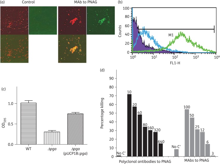
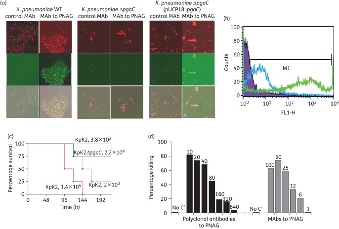
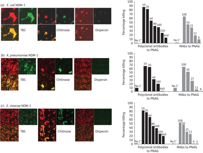
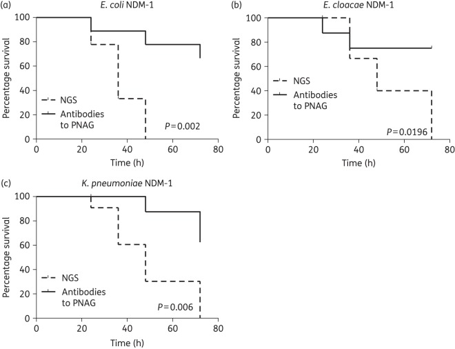
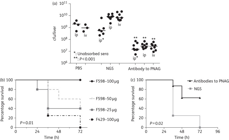
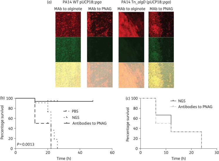
Methods: PNAG production was detected by confocal microscopy and its role in the formation of the biofilm (for E. cloacae) and as a virulence factor (for K. pneumoniae) was analysed. The in vitro (opsonophagocytosis killing assay) and in vivo (mouse models of peritonitis) activity of antibodies to PNAG were studied using antibiotic-susceptible and -resistant E. coli, E. cloacae and K. pneumoniae. A PNAG-producing strain of Pseudomonas aeruginosa, an organism that does not naturally produce this antigen, was constructed by adding the pga locus to a strain with inactive alg genes responsible for the production of P. aeruginosa alginate. Antibodies to PNAG were tested in vitro and in vivo as above.

Results: PNAG is a major component of the E. cloacae biofilm and a virulence factor for K. pneumoniae. Antibodies to PNAG mediated in vitro killing (>50%) and significantly protected mice against the New Delhi metallo-β-lactamase-producing E. coli (P = 0.02), E. cloacae (P = 0.0196) and K. pneumoniae (P = 0.006), against K. pneumoniae carbapenemase (KPC)-producing K. pneumoniae (P = 0.02) and against PNAG-producing P. aeruginosa (P = 0.0013). Thus, regardless of the Gram-negative bacterial species, PNAG expression is the sole determinant of the protective efficacy of antibodies to this antigen.

Conclusions: Our findings suggest antibodies to PNAG may provide extended-spectrum antibacterial protective activity.


Figures

Figure 1.
Figure 1.
E. cloacae and PNAG production. Detection of PNAG on the surface of E. cloacae 2a using immunofluorescence confocal microscopy (a) and flow cytometry (b). Binding of MAb F598 to PNAG conjugated to AF488 [green fluorescence in (a) and green curve in (b)]. No binding with the control MAb conjugated to AF488 was detected [lack of green fluorescence in (a) and blue curve in (b)]. In (a), SYTO 83 was used to visualize DNA (red fluorescence) and an overlay of red and green channels is presented. (c) Formation of biofilms after 2 days of growth at room temperature comparing E. cloacae 2a, E. cloacae 2a Δpga and E. cloacae 2a Δpga (pUCP18::pga). (d) Opsonophagocytic killing of E. cloacae 2a mediated by polyclonal antibodies and MAbs to PNAG. Bars represent mean percentage of killing relative to the control containing NGS or the control MAb F429. All standard deviations (not shown) were <15%. Assays were done in duplicate. C′, complement; absence of killing arbitrarily assigned as 1% killing. Dilutions of polyclonal antibodies and amounts (in μg) of MAb to PNAG tested are provided at the top of each bar.
Figure 2.
Figure 2.
K. pneumoniae and PNAG production. Detection of PNAG on the surface of K. pneumoniae K2. (a) Immunofluorescence confocal microscopy. (b) Flow cytometry analyses. Binding of MAb F598 to PNAG conjugated to AF488 [green fluorescence in (a) and green curve in (b)]. No binding of the control MAb [lack of green fluorescence in (a) and blue curve in (b)]. In (a), SYTO 83 was used to visualize DNA (red fluorescence). Bacteria tested in (a): K. pneumoniae K2, K. pneumoniae K2 ΔpgaC and K. pneumoniae K2 ΔpgaC (pUCP18::pgaC) (c). Impact of loss of PNAG on the virulence of K. pneumoniae K2: C3H/H3N mice (8/group) were challenged intraperitoneally with K. pneumoniae K2 (KpK2) at doses of 1.4 × 104, 2 × 103 or 3.8 × 102 cfu/mouse or with 2.2 × 104 cfu of K. pneumoniae K2 ΔpgaC (KpK2ΔpgaC). (d) Opsonophagocytic killing of K. pneumoniae K2 mediated by polyclonal antibodies or MAbs to PNAG. Bars indicate mean percentage of killing relative to controls containing NGS or the control MAb F429. All standard deviations (not shown) were <13%. Assays were done in duplicate. C′, complement; absence of killing arbitrarily assigned as 1% killing. Dilutions of polyclonal antibodies and amounts (in μg) of MAbs to PNAG tested are provided at the top of each bar.
Figure 3.
Figure 3.
PNAG production by NDM-1-producing Enterobacteriaceae and in vitro killing by PNAG antibodies. Detection of PNAG on the surface of NDM-1-producing Enterobacteriaceae strains using immunofluorescence confocal microscopy (left panels). Binding of MAb F598 to PNAG conjugated to AF488 results in green fluorescence. SYTO 83 was used to visualize DNA (red fluorescence). Bacteria tested: NDM-1-producing E. coli, K. pneumoniae and E. cloacae (a, b, c left panels, respectively). Digestion with the PNAG-degrading enzyme dispersin B, but not a control enzyme, chitinase, resulted in loss of binding of MAb F598. Opsonophagocytic killing of NDM-1-producing E. coli, K. pneumoniae and E. cloacae (a, b, c, right panels, respectively) mediated by polyclonal antibodies or MAbs to PNAG. Bars represent mean percentage of killing relative to control containing NGS or the control MAb F429. All standard deviations (not shown) were <15%. Assays were done in duplicate. C′, complement; absence of killing arbitrarily assigned as 1% killing. Dilutions of polyclonal antibodies and concentrations (in μg) of MAbs to PNAG tested are provided at the top of each bar.
Figure 4.
Figure 4.
In vivo protection by polyclonal antibodies to PNAG against E. coli NDM-1, E. cloacae NDM-1 and K. pneumoniae NDM-1 infections. C3H/H3N mice (8/group) were passively immunized with PNAG-specific goat antiserum intraperitoneally 24 and 4 h before intraperitoneal infection with E. coli NDM-1 (108 cfu/mouse), E. cloacae NDM-1 (5 × 109 cfu/mouse) or K. pneumoniae NDM-1 (5 × 108 cfu/mouse). Controls received NGS. P values by log-rank test.
Figure 5.
Figure 5.
In vivo protection by MAbs and polyclonal antibodies to PNAG against K. pneumoniae KPC infections. (a) C3H/H3N mice (n = 24) were passively immunized with PNAG-specific goat antiserum given intraperitoneally (ip) or intravenously (iv) 24 h and 4 h before intraperitoneal inoculation of K. pneumoniae KPC. Controls received NGS (n = 20) or PBS (n = 7). All sera were either intact or absorbed with K. pneumoniae K2ΔpgaC to remove antibodies to K. pneumoniae other than those to PNAG. The challenge dose was 108 cfu/mouse. P values by unpaired non-parametric t-test comparing NGS to immune serum in mice challenged by the same route. (b) C3H/H3N mice (8/group) were passively immunized with different amounts of the MAb to PNAG (from 25 to 100 μg) or MAb to P. aeruginosa alginate (100 μg) as a control then challenged intravenously with K. pneumoniae KPC (109 cfu/mouse). (c) Mice were injected intraperitoneally with PNAG-specific goat antiserum 24 and 4 h before inoculation with K. pneumoniae KPC. Controls received NGS. P values by log-rank test comparing MAb F598 (100 μg) with MAb F429 (100 μg) (b) or antibodies to PNAG to non-immune serum (c).
Figure 6.
Figure 6.
Impact of PNAG production in P. aeruginosa on protective efficacy of antibody to PNAG. (a) Detection of PNAG on the surface of P. aeruginosa. Binding of MAb F598 to PNAG conjugated to AF488 results in green fluorescence. SYTO 83 was used to visualize DNA (red fluorescence). The overlap of the green and red colour is presented in the bottom of each panel. WT P. aeruginosa PA14 carrying a cloned pga locus [PA14 WT (pUCP18::pga)] was positive for alginate production, but negative for PNAG. P. aeruginosa PA14 with a Tn insertion in the algD gene to inactivate alginate production (PA14 Tn::algD) with a cloned pga locus [PA14 Tn::algD (pUCP18::pga)] was negative for alginate, but positive for PNAG production. (b) C3H/H3N mice were passively immunized with PBS (4 mice), NGS (6 mice) or PNAG-specific goat immune serum (6 mice) intraperitoneally 24 and 4 h before intraperitoneal challenge with PNAG-producing P. aeruginosa PA14 Tn::algD (pUCP18::pga) (4 × 107 cfu/mouse). (c) C3H/H3N mice (n = 8/group) were passively immunized with either NGS or PNAG-specific goat immune serum intraperitoneally 24 and 4 h before intraperitoneal challenge with PNAG- and alginate-negative P. aeruginosa PA14 Tn::algD (5 × 108 cfu/mouse). P values by log-rank test comparing NGS with antibodies with PNAG.

Similar articles

Cited by

References

    1. McKenney D, Hubner J, Muller E et al. . The ica locus of Staphylococcus epidermidis encodes production of the capsular polysaccharide/adhesin. Infect Immun 1998; 66: 4711–20. - PMC - PubMed
    1. Wang X, Preston JF 3rd, Romeo T. The pgaABCD locus of Escherichia coli promotes the synthesis of a polysaccharide adhesin required for biofilm formation. J Bacteriol 2004; 186: 2724–34. - PMC - PubMed
    1. Cywes-Bentley C, Skurnik D, Zaidi T et al. . Antibody to a conserved antigenic target is protective against diverse prokaryotic and eukaryotic pathogens. Proc Natl Acad Sci USA 2013; 110: E2209–18. - PMC - PubMed
    1. Skurnik D, Davis MR Jr, Benedetti D et al. . Targeting pan-resistant bacteria with antibodies to a broadly conserved surface polysaccharide expressed during infection. J Infect Dis 2012; 205: 1709–18. - PMC - PubMed
    1. Cerca N, Maira-Litran T, Jefferson KK et al. . Protection against Escherichia coli infection by antibody to the Staphylococcus aureus poly-N-acetylglucosamine surface polysaccharide. Proc Natl Acad Sci USA 2007; 104: 7528–33. - PMC - PubMed

Publication types

MeSH terms