. 2016 Mar;170(3):1255-70.
doi: 10.1104/pp.15.01232. Epub 2016 Jan 8.

Deficiency of Starch Synthase IIIa and IVb Alters Starch Granule Morphology from Polyhedral to Spherical in Rice Endosperm

Affiliations

Deficiency of Starch Synthase IIIa and IVb Alters Starch Granule Morphology from Polyhedral to Spherical in Rice Endosperm

Yoshiko Toyosawa et al. Plant Physiol. 2016 Mar.

Abstract

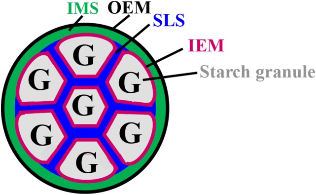
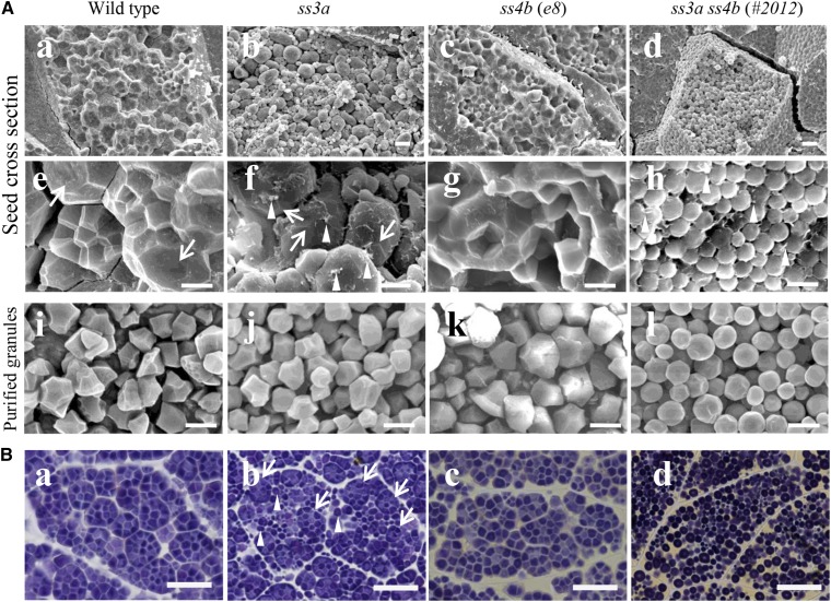
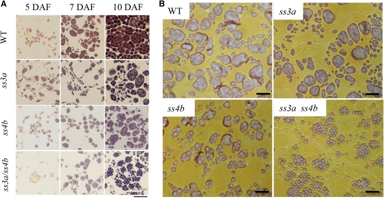
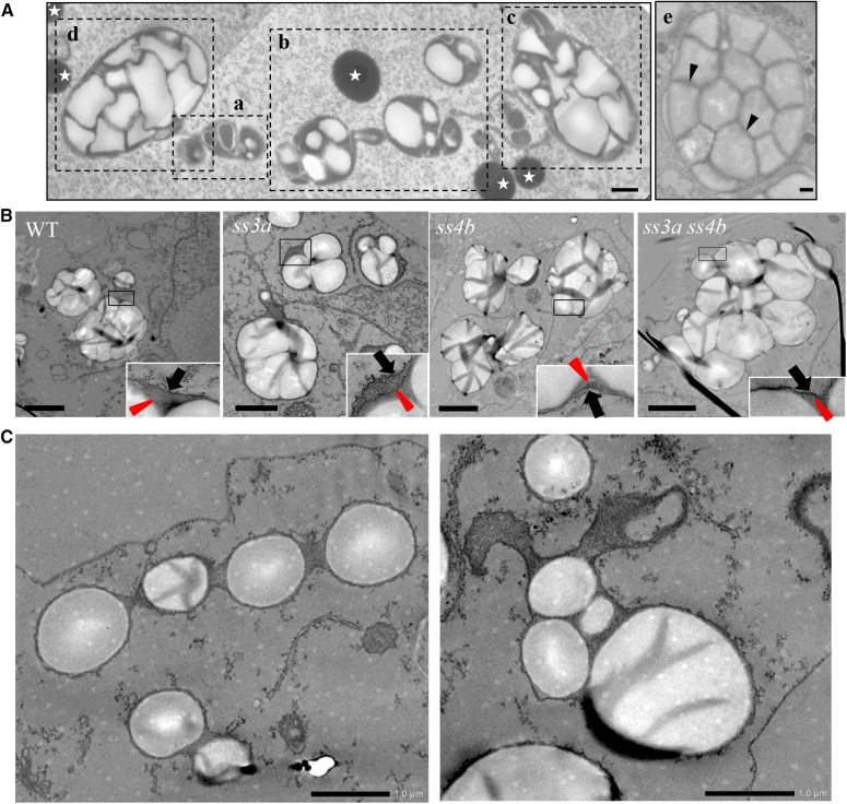
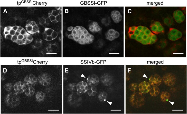
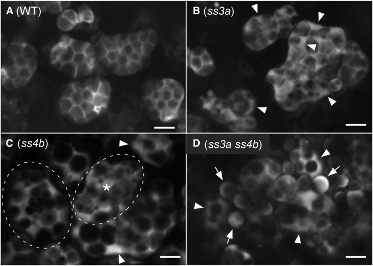
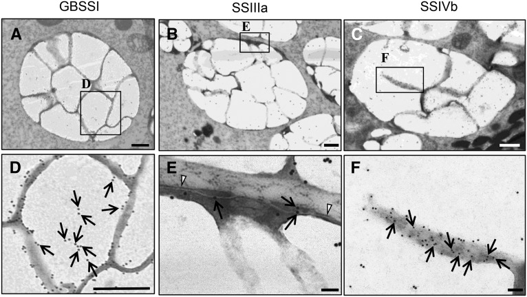
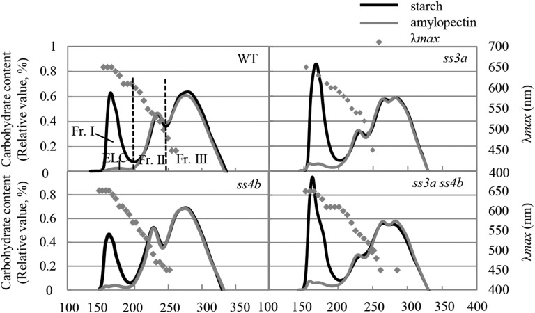
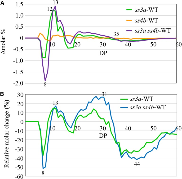
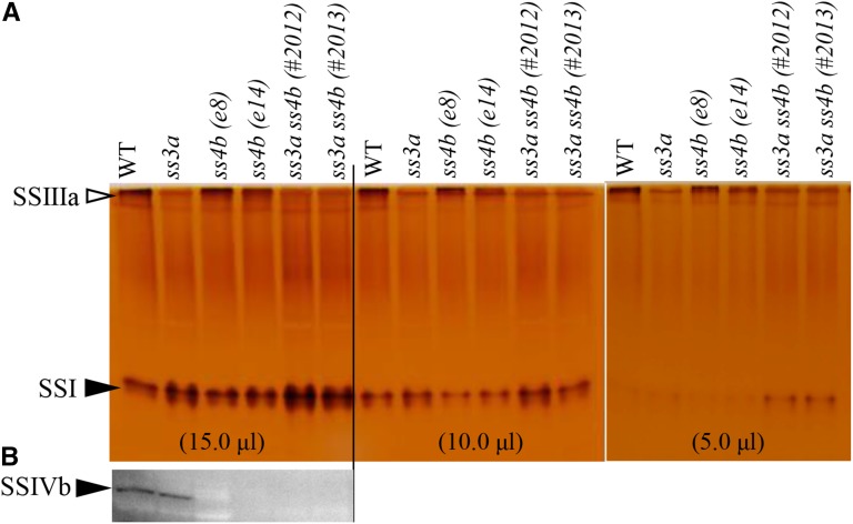
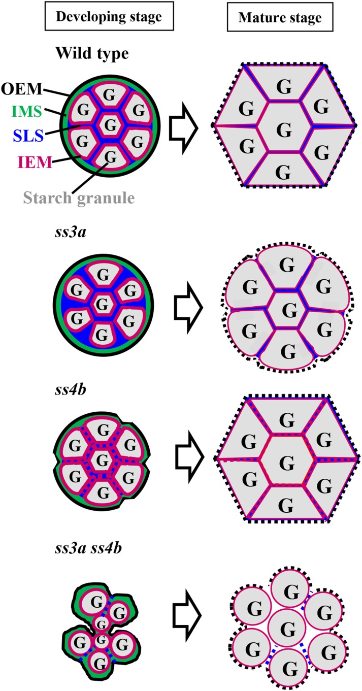
Starch granule morphology differs markedly among plant species. However, the mechanisms controlling starch granule morphology have not been elucidated. Rice (Oryza sativa) endosperm produces characteristic compound-type granules containing dozens of polyhedral starch granules within an amyloplast. Some other cereal species produce simple-type granules, in which only one starch granule is present per amyloplast. A double mutant rice deficient in the starch synthase (SS) genes SSIIIa and SSIVb (ss3a ss4b) produced spherical starch granules, whereas the parental single mutants produced polyhedral starch granules similar to the wild type. The ss3a ss4b amyloplasts contained compound-type starch granules during early developmental stages, and spherical granules were separated from each other during subsequent amyloplast development and seed dehydration. Analysis of glucan chain length distribution identified overlapping roles for SSIIIa and SSIVb in amylopectin chain synthesis, with a degree of polymerization of 42 or greater. Confocal fluorescence microscopy and immunoelectron microscopy of wild-type developing rice seeds revealed that the majority of SSIVb was localized between starch granules. Therefore, we propose that SSIIIa and SSIVb have crucial roles in determining starch granule morphology and in maintaining the amyloplast envelope structure. We present a model of spherical starch granule production.


Figures

Figure 1.
Figure 1.
Structural model of the wild-type amyloplast in developing rice endosperm. The OEM is in black, the IEM is in magenta, the IMS is in green, and the SLS is in blue. G, Starch granules.
Figure 2.
Figure 2.
Starch granule morphology in mature rice seeds. A, Scanning electron micrographs of cross sections of mature seeds (central part of the endosperm; a–h) and purified starch granules (i–l). Arrows indicate the amyloplast surface, and arrowheads indicate putative shreds of the amyloplast envelope. Bars = 10 μm (a–d) and 5 μm (e–l). B, Light microscopy of iodine-stained thin cross sections of mature seeds (central part of the endosperm). Arrows indicate compound-type granules, and arrowheads indicate spherical granules. Bars = 20 μm.
Figure 3.
Figure 3.
Starch granule morphology in developing endosperm. Light microscopy shows thin iodine-stained sections of the central part of the developing endosperm. Wild-type cv Nipponbare (WT), ss3a, ss4b (e8), and ss3a ss4b (#2012) were compared. A, Developing endosperm at 5, 7, and 10 DAF. Bar = 20 μm. B, Developing endosperm at 7 DAF. Bars = 10 μm.
Figure 4.
Figure 4.
Transmission electron micrographs showing amyloplast morphology in developing rice starchy endosperm. A, Amyloplast development in the subaleurone layer cells of the wild type. a to e show amyloplasts at different developmental stages. Arrowheads indicate the IEM, and stars show protein bodies. Bars = 500 nm. B, Typical amyloplasts observed in ultrathin uranyl-lead-stained sections in the subaleurone layer cells at 7 DAF. Wild-type cv Nipponbare (WT), ss3a, ss4b (e8), and ss3a ss4b (#2012) were compared. Arrowheads indicate IEM, and arrows indicate outer amyloplast membrane. Bars = 2 μm. C, Aberrant ss3a ss4b amyloplasts in the subaleurone layer cells at 7 DAF. Bars = 1 μm.
Figure 5.
Figure 5.
Distinct localizations of GBSSI and SSIVb. Confocal fluorescence microscopy shows developing seeds of transformed wild-type (cv Yukihikari) plants (7 DAF) expressing tpGBSSICherry together with either GBSSI-GFP (A–C) or SSIVb-GFP (D–F). A to C, tpGBSSICherry (magenta channel) and GBSSI-GFP (green channel). Note that the two signals do not overlap, indicating that tpGBSSICherry is not efficiently imported into the stroma but is located primarily in the SLS and envelope. D to F, tpGBSSICherry (magenta channel) and SSIVb-GFP (green channel). Note that the SSIVb-GFP and tpGBSSICherry signals overlap extensively and that SSIVb-GFP is also detected as dots (arrowheads). Bars = 5 μm.
Figure 6.
Figure 6.
Internal structures of amyloplasts visualized with tpGBSSIGFP. Developing seeds of the transformed wild-type (WT) cv Nipponbare (A), ss3a (B), ss4b (e14; C), and ss3a ss4b (#2013; D) were analyzed at 7 DAF by confocal microscopy. In B and C, arrowheads indicate enlarged IMS between the OEM and the IEM. In C, an amyloplast indicated by a dashed oval and asterisk does not contain a typical SLS lattice structure. Another amyloplast, indicated by a dashed oval, contains a cluster of small granules in the center and large granules at the periphery. Note that these distinct phenotypes in ss4b were observed with low frequency (less than 10%). Most amyloplast phenotypes were similar to that of the wild type. In D, arrowheads indicate amyloplasts containing multiple starch granules and arrows indicate granules that look like simple-type grains. Bars = 5 μm.
Figure 7.
Figure 7.
Immunoelectron microscopy of amyloplasts and the distribution of GBSSI, SSIIIa, and SSIVb in developing endosperm (7 DAF) of wild-type rice (cv Taichung 65). The distributions of GBSSI (A and D), SSIIIa (B and E), and SSIVb (C and F) are indicated by 15-nm gold particles (arrows). Arrowheads indicate the OEM. Bars = 500 nm (A–C) and 100 nm (D–F).
Figure 8.
Figure 8.
Gel filtration chromatography of isoamylase-debranched starch and purified amylopectin. Isoamylase-debranched starch (black lines) and purified amylopectin (gray lines) from wild-type cv Nipponbare (WT), ss3a, ss4b (e8), and ss3a ss4b (#2012) were analyzed by gel filtration chromatography on a series of Toyopearl HW55S and Toyopearl HW50S columns. The proportions of the starch components calculated from these data are shown in Supplemental Table S4. ELC, Extra-long chain; Fr., fraction.
Figure 9.
Figure 9.
Chain length distribution patterns of amylopectin in wild-type rice and mutant lines. A, Differences in the chain length distribution patterns (Δmolar%) between mutant lines and wild-type cv Nipponbare (WT). Values represent averages of three seeds arbitrarily chosen from a single homozygous plant. B, Relative molar changes of each amylopectin chain (Δmol %/mol % × 100) calculated from the data shown in A for DP 6 to 60 for the wild type, ss3a, and ss3a ss4b (#2012). The numbers on the plots are DP values.
Figure 10.
Figure 10.
Detection of SS activity (SSI and SSIIIa) and SSIVb protein. Numbers in parentheses represent the volumes of the crude enzyme extract per lane. A, Native PAGE/SS activity staining. SSIIIa and SSI activity bands are indicated by arrowheads. B, Immunoblotting using antiserum against rice SSIVb. WT, Wild-type cv Nipponbare.
Figure 11.
Figure 11.
Possible molecular mechanism underlying amyloplast and starch granule synthesis in wild-type rice and mutant lines. Amyloplast development is shown in wild-type rice and in ss3a, ss4b, and ss3a ss4b mutants. At left are amyloplasts during early seed development (7 DAF), and at right are amyloplasts in mature seeds. The black dashed lines in mature seeds show the disrupted OEM during seed maturation and/or desiccation.

References

    1. Ball S, Colleoni C, Cenci U, Raj JN, Tirtiaux C (2011) The evolution of glycogen and starch metabolism in eukaryotes gives molecular clues to understand the establishment of plastid endosymbiosis. J Exp Bot 62: 1775–1801 - PubMed
    1. Crumpton-Taylor M, Pike M, Lu KJ, Hylton CM, Feil R, Eicke S, Lunn JE, Zeeman SC, Smith AM (2013) Starch synthase 4 is essential for coordination of starch granule formation with chloroplast division during Arabidopsis leaf expansion. New Phytol 200: 1064–1075 - PMC - PubMed
    1. Delvallé D, Dumez S, Wattebled F, Roldán I, Planchot V, Berbezy P, Colonna P, Vyas D, Chatterjee M, Ball S, et al. (2005) Soluble starch synthase I: a major determinant for the synthesis of amylopectin in Arabidopsis thaliana leaves. Plant J 43: 398–412 - PubMed
    1. Deschamps P, Colleoni C, Nakamura Y, Suzuki E, Putaux JL, Buléon A, Haebel S, Ritte G, Steup M, Falcón LI, et al. (2008) Metabolic symbiosis and the birth of the plant kingdom. Mol Biol Evol 25: 536–548 - PubMed
    1. Fujita N, Hasegawa H, Taira T (2001) The isolation and characterization of a waxy mutant of diploid wheat (Triticum monococcum L.). Plant Sci 160: 595–602 - PubMed

Publication types

MeSH terms

LinkOut - more resources