. 2016 Feb;18(1):4.
doi: 10.1007/s10544-015-0027-x.

Facile and cost-effective production of microscale PDMS architectures using a combined micromilling-replica moulding (μMi-REM) technique

Affiliations

Facile and cost-effective production of microscale PDMS architectures using a combined micromilling-replica moulding (μMi-REM) technique

Dario Carugo et al. Biomed Microdevices. 2016 Feb.

Abstract

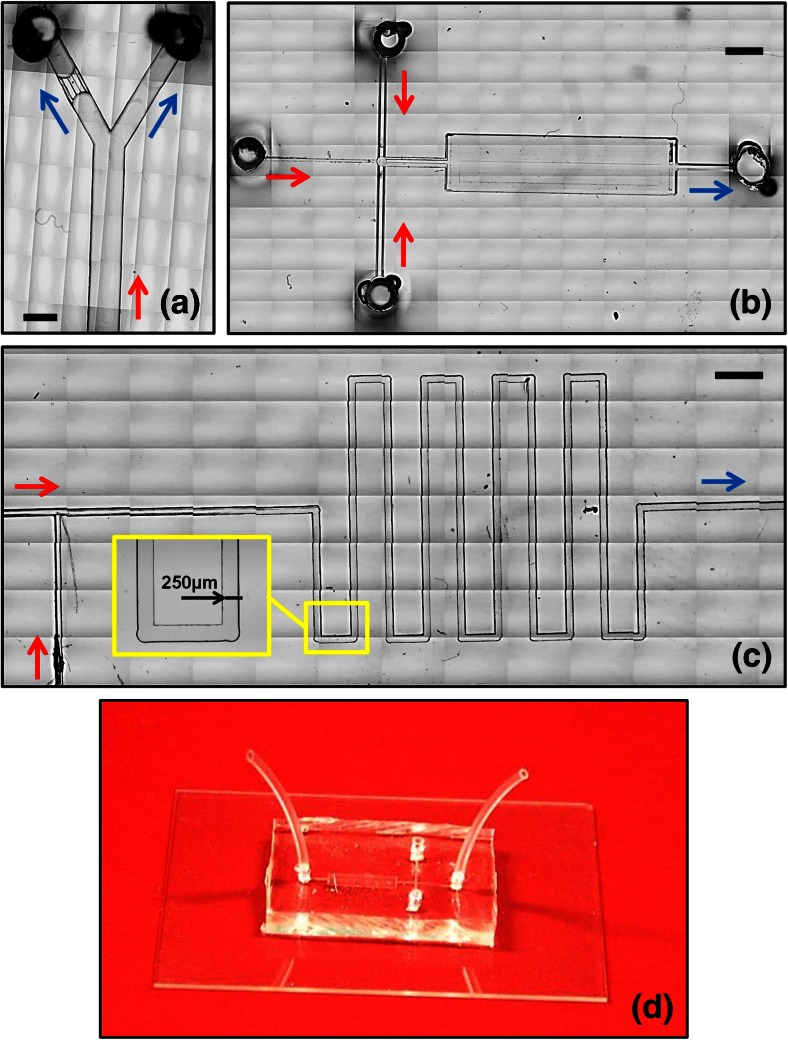
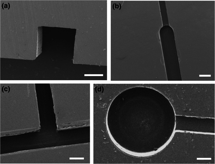
We describe a cost-effective and simple method to fabricate PDMS-based microfluidic devices by combining micromilling with replica moulding technology. It relies on the following steps: (i) microchannels are milled in a block of acrylic; (ii) low-cost epoxy adhesive resin is poured over the milled acrylic block and allowed to cure; (iii) the solidified resin layer is peeled off the acrylic block and used as a mould for transferring the microchannel architecture onto a PDMS layer; finally (iv) the PDMS layer is plasma bonded to a glass surface. With this method, microscale architectures can be fabricated without the need for advanced technological equipment or laborious and time-consuming intermediate procedures. In this manuscript, we describe and validate the microfabrication procedure, and we illustrate its applicability to emulsion and microbubble production.

Keywords: Emulsions; Microbubbles; Microchannel; Microfabrication; Microfluidic; Micromilling; Pdms; Pmma; Replica moulding.


Figures

Fig. 1
Fig. 1
Temporal evolution of the number of scientific publications containing the keywords “PDMS + microfluidic” from Google Scholar (empty black squares) and Elsevier Scopus® (empty red circles) from 2000 to 2014
Fig. 2
Fig. 2
Microdevice fabrication by combined micromilling and replica moulding (μMi-REM)
Fig. 3
Fig. 3
Imaging of channel cross-section using scanning electron microscopy (SEM) and measurement of channel width (w) and height (h) from the acquired microscope images. These were quantified for each moulding step. For the representative PDMS microchannel reported here, h = 117.8 μm and w = 272.6 μm
Fig. 4
Fig. 4
a-c Bright-field microscope images of microfluidic devices fabricated using μMi-REM. The images were created from a stack of multiple microscope acquisitions over a large surface area of the device, acquired using a Nikon ECLIPSE Ti inverted microscope (Nikon Corporation, Tokyo, Japan) coupled with a charge-coupled device (CCD) camera (Digital sight Ds-Fi1, Nikon Corporation, Tokyo, Japan). The red and blue arrows indicate the inflow and outflow lines, respectively. The inlet and outlet ports in (a) and (b) were fabricated using the method reported in Supplementary Section S2. a Single bifurcation architecture, with a ‘micro-filter’ structure on the left daughter branch (microchannels are 500 μm deep). b Cross-flow architecture followed by a micro-chamber for visualization purposes (microchannels are 50 μm deep). c T-junction architecture followed by a serpentine-like structure. A magnified view of a serpentine section is shown in the inset (microchannels are 50 μm deep). d Finalised microfluidic device, with the PDMS layer containing the microchannel architecture plasma bonded to a 1 mm thick glass layer. Inlet and outlet tubing can be directly connected to the inlet/outlet ports of the device (through pre-formed reservoirs), without the need for additional connection elements. Scale bars in (a), (b), (c) are 500 μm, 2 mm, and 1 mm, respectively.
Fig. 5
Fig. 5
Representative SEM images of PDMS microchannels fabricated using μMi-REM. Scale bars are equal to 250 μm (a), 125 μm (b), 500 μm (c), and 200 μm (d)
Fig. 6
Fig. 6
(a) Schematic of the ‘T-junction’ microfluidic device employed for the production of emulsions and microbubbles. Channel IN1 is 127 μm wide and 50 μm deep, whilst channels IN2 and OUT are 254 μm wide and 50 μm deep. The inset shows the formation of PBS-in-PLGA emulsions (stained using Evans blue) in a region of the device located after the T-junction. b-c Bright-field microscope images of PBS-in-PLGA emulsions (stained with Evans blue) (b) and phospholipid-shelled microbubbles (c). Images (magnification: 4×) were acquired using a Leica DM500 microscope (Leica Microsystems GmbH, Wetzlar, Germany) coupled with a CCD camera (MicroPublisher 3.3 RTV, QImaging, Surrey, Canada). (d) Representative size distribution of microbubbles obtained from a single experimental run (total number of counted bubbles =184). A population of bubbles with radius lower than 120 μm is present (corresponding to less than 8 % of the total bubble population) and is likely to be attributed to flow fluctuations originating from the stepped motor of the syringe pump

References

    1. Ashley JF, Cramer NB, et al. Soft-lithography fabrication of microfluidic features using thiol-ene formulations. Lab Chip. 2011;11(16):2772–2778. doi: 10.1039/c1lc20189a. - DOI - PubMed
    1. Becker H, Heim U. Hot embossing as a method for the fabrication of polymer high aspect ratio structures. Sensors Actuators A Phys. 2000;83(1):130–135. doi: 10.1016/S0924-4247(00)00296-X. - DOI
    1. Borenstein JT, Terai H, et al. Microfabrication technology for vascularized tissue engineering. Biomed. Microdevices. 2002;4(3):167–175. doi: 10.1023/A:1016040212127. - DOI
    1. Borenstein JT, Tupper MM, et al. Functional endothelialized microvascular networks with circular cross-sections in a tissue culture substrate. Biomed. Microdevices. 2010;12(1):71–79. doi: 10.1007/s10544-009-9361-1. - DOI - PubMed
    1. Bose N, Das T, et al. Enhancement of static incubation time in microfluidic cell culture platforms exploiting extended air–liquid interface. Lab Chip. 2012;12(1):69–73. doi: 10.1039/C1LC20888H. - DOI - PubMed

Publication types

MeSH terms

Substances