. 2016 Jul;100(1):185-94.
doi: 10.1189/jlb.3A0515-185R. Epub 2016 Jan 8.

Mincle suppresses Toll-like receptor 4 activation

Affiliations

Mincle suppresses Toll-like receptor 4 activation

Stephanie H Greco et al. J Leukoc Biol. 2016 Jul.

Abstract

Regulation of Toll-like receptor responses is critical for limiting tissue injury and autoimmunity in both sepsis and sterile inflammation. We found that Mincle, a C-type lectin receptor, regulates proinflammatory Toll-like receptor 4 signaling. Specifically, Mincle ligation diminishes Toll-like receptor 4-mediated inflammation, whereas Mincle deletion or knockdown results in marked hyperresponsiveness to lipopolysaccharide in vitro, as well as overwhelming lipopolysaccharide-mediated inflammation in vivo. Mechanistically, Mincle deletion does not up-regulate Toll-like receptor 4 expression or reduce interleukin 10 production after Toll-like receptor 4 ligation; however, Mincle deletion decreases production of the p38 mitogen-activated protein kinase-dependent inhibitory intermediate suppressor of cytokine signaling 1, A20, and ABIN3 and increases expression of the Toll-like receptor 4 coreceptor CD14. Blockade of CD14 mitigates the increased sensitivity of Mincle(-/-) leukocytes to Toll-like receptor 4 ligation. Collectively, we describe a major role for Mincle in suppressing Toll-like receptor 4 responses and implicate its importance in nonmycobacterial models of inflammation.

Keywords: C-type lectin receptor; inflammation; sepsis.


Figures

Figure 1
Figure 1
Mincle deletion results in exaggerated TLR4 responses. (A) Splenocytes derived from WT or Mincle−/− mice (n = 3) were stimulated with TLR4 ligand (LPS, 10 µg/ml) and tested for expression of activating cytokines in cell culture supernatant after 24 h. (B) CD11c+ BMDCs derived from WT or Mincle−/− mice were stimulated with PBS or LPS and tested for expression of TNF‐α by intracellular cytokine analysis. (C–E) WT and Mincle−/− BMDCs were stimulated with PBS or LPS and tested for expression of TNF‐α (C), IL‐6 (D), and MCP‐1 (E) in cell culture supernatant after 24 h. (F–H) WT or Mincle−/− BMDMs were stimulated with PBS or LPS and tested for expression of TNF‐α (F), IL‐6 (G), and MCP‐1 (H). Experiments were performed >3 times in triplicate. *P < 0.05.
Figure 2
Figure 2
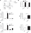
Mincle inhibition or knockdown results in exaggerated TLR4 responses. (A) Splenocytes derived from WT or Mincle−/− mice were stimulated with TLR4 ligand (LPS, 10µg/ml) after pretreatment with a neutralizing Mincle mAb (5 µg/ml) or isotype control and tested for expression of TNF‐α in cell culture supernatant at 24 h. (B and C) Knockdown of Mincle in Raji cells was confirmed by flow cytometry based on mean fluorescence intensity (B) and Western blotting (C). Density analysis based on triplicates is shown. (D and E) Raji cell siRNA Mincle transfectants and controls were tested for expression of IL‐6 (D) and IL‐8 (E) in 24‐h cell culture supernatant after stimulation with LPS. Experiments were repeated 3 times with similar results. *P < 0.05, **P < 0.01.
Figure 3
Figure 3
Mincle−/− mice are more susceptible to endotoxic shock. WT and Mincle−/− mice were treated with LPS i.p. and (A–D) tested for serum levels of proinflammatory cytokines at timed intervals. Splenic macrophages (E), dendritic cells (F), inflammatory monocytes (G), and neutrophils (H) were tested for expression of TNF‐α at 6 h and 24 h by intracellular cytokine analysis. (I) The changes in core body temperature from baseline in LPS‐treated mice were measured using a rectal probe. *P < 0.05, **P < 0.01, ***P < 0.001. (J) Survival was assessed according to the Kaplan‐Meier method (n = 5 mice per group; P < 0.001). In vivo experiments were repeated twice with similar results. H, hours.
Figure 4
Figure 4
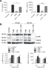
Mincle ligation inhibits TLR4 activation. (A) Splenocytes were stimulated with LPS (100 ng/ml), TDB, or costimulated with LPS + TDB and tested for expression of IL‐6 and TNF‐α in cell culture supernatant after 24 h. (B) Cells were similarly stimulated with LPS and TDB, alone or in combination, for the indicated time intervals and tested for expression of β‐actin, TRAF6, and p‐JNK by Western blotting. Density plots based on triplicates are shown. Experiments were repeated 3 times with similar results. *P < 0.05, ***P < 0.001.
Figure 5
Figure 5
Mincle modulates expression of proinflammatory and inhibitory signaling pathways. (A and B) WT or Mincle−/− splenocytes were stimulated with LPS for timed intervals and tested for expression of p‐ERK, ERK, p‐JNK, and TRAF6 (A) and phosphorylated and nonphosphorylated Syk and p38, SOCS1, A20, ABIN3, Mal, SHP‐1, Hes‐1, and TANK (B) by Western blotting. β‐actin was used as a loading control. (C) Density plots based on triplicates are shown. *P < 0.05.
Figure 6
Figure 6
Mincle deletion does not suppress IL‐10 expression or alter its inhibitory function after LPS stimulation. BMDC (A), BMDM (B), and splenocyte (C) concentrates from WT or Mincle−/− mice were stimulated with LPS (10 µg/ml) and tested for IL‐10 production in culture supernatant after 24 h. (D) WT and Mincle−/− mice were injected with LPS i.p (10 mg/kg), and serum IL‐10 levels were measured at serial time intervals (n = 3/group). (E) WT and Mincle−/− BMDCs were pretreated in culture with PBS or recombinant IL‐10 (10 ng/ml) 1 h before overnight stimulation with PBS or LPS (100 ng/ml). Cell culture supernatant was tested for IL‐6. Each experiment was repeated at least twice in triplicates with similar results. ns, not significant; *P < 0.05.
Figure 7
Figure 7
Mincle and TLR4 reciprocally cross‐regulate each other after receptor ligation. (A) WT BMDM were treated with PBS or TDB and tested for TLR4 expression. Mean fluorescence intensities (MFIs) are shown. (B) Similarly, BMDMs were stimulated with PBS or LPS (10 µg/ml) and tested for Mincle expression. (C) Expression of TLR4 was tested in splenic dendritic cells, macrophages, and neutrophils from WT and Mincle−/− mice. Representative data and average MFIs from experiments repeated 3 times are shown. ns, not significant; *P < 0.05; **P < 0.01.
Figure 8
Figure 8
Mincle regulates TLR coreceptor expression. BMDMs (A) or splenocyte suspensions (B) derived from WT or Mincle−/− mice were treated with PBS or LPS (10 µg/ml) and tested for expression of CD14 by flow cytometry (A) (fluorescence intensities are shown) and PCR (B). (C and D) Splenocytes derived from WT and Mincle−/− mice were treated with PBS or a neutralizing CD14 mAb (10 µg/ml) and LPS, either alone, or in combination. Expression of TNF‐α (C) and IL‐6 (D) were tested in cell culture supernatant. The relative decrease in cytokine production upon CD14 blockade in Mincle−/− leukocytes compared with WT leukocytes was calculated. Experiments were repeated twice with similar results. ***P < 0.001.

References

    1. Akira, S. , Takeda, K. (2004) Toll‐like receptor signalling. Nat. Rev. Immunol. 4, 499–511. - PubMed
    1. Kawai, T. , Akira, S. (2010) The role of pattern‐recognition receptors in innate immunity: update on Toll‐like receptors. Nat. Immunol. 11, 373–384. - PubMed
    1. Bianchi, M. E. (2007) DAMPs, PAMPs and alarmins: all we need to know about danger. J. Leukoc. Biol. 81, 1–5. - PubMed
    1. Kondo, T. , Kawai, T. , Akira, S. (2012) Dissecting negative regulation of Toll‐like receptor signaling. Trends Immunol. 33, 449–458. - PubMed
    1. Takeuchi, O. , Kawagoe, T. , Akira, S. (2009) Tank is a negative regulator of TLR signaling and critical for preventing autoimmune nephritis. Cytokine 48, 20. - PMC - PubMed

Publication types

MeSH terms