

Periventricular hyperintensities are associated with elevated cerebral amyloid
- PMID: 26747881
- PMCID: PMC4753726
- DOI: 10.1212/WNL.0000000000002352
Periventricular hyperintensities are associated with elevated cerebral amyloid
Abstract
Objective: To investigate the association between periventricular white mater hyperintensities (PVWMH) and biomarkers of elevated cerebral β-amyloid (Aβ) in the Alzheimer's Disease Neuroimaging Initiative, a large prospective multicenter observational study.
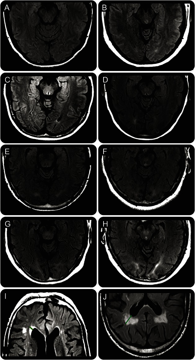
Methods: The burden of frontal, parietal, and occipital PVWMH on 3T fluid-attenuated inversion recovery MRI was evaluated in 698 cognitively normal participants and participants with mild cognitive impairment (MCI) using a novel semiquantitative visual rating scale. Results were correlated with CSF-Aβ, florbetapir-PET, and fluorodeoxyglucose (FDG)-PET.
Results: Increased burden of parietal, occipital, and frontal PVWMH was associated with elevated cerebral amyloid evidenced by high florbetapir-PET signal (p < 0.01) and low CSF-Aβ (p < 0.01). In logistic regression models, including PVWMH, age, sex, APOE status, vascular risk factors, pulse pressure, vascular secondary prevention medications, education, ethnicity, and race, parietal, occipital, and frontal PVWMH burden was independently associated with high florbetapir-PET uptake (p < 0.05). In a similar logistic regression model, parietal and occipital (p < 0.05) but not frontal (p = 0.05) PVWMH were independently associated with CSF-Aβ. Weaker associations were found between parieto-occipital PVWMH and elevated CSF-tau (p < 0.05) and occipital PVWMH and elevated CSF-phospho-tau (p < 0.05). PVWMH were associated with cerebral hypometabolism on FDG-PET independent of CSF-Aβ levels (p < 0.05). Absolute and consistency of agreement intraclass correlation coefficients were, respectively, 0.83 and 0.83 for frontal, 0.78 and 0.8 for parietal, and 0.45 and 0.75 for occipital PVWMH measurements.
Conclusions: Increased PVWMH were associated with elevated cerebral amyloid independent of potential confounders such as age, APOE genotype, and vascular risk factors. The mechanisms underlying the association between PVWMH and cerebral amyloid remain to be clarified.
© 2016 American Academy of Neurology.
Figures
References
-
- Thal DR, Attems J, Ewers M. Spreading of amyloid, tau, and microvascular pathology in Alzheimer's disease: findings from neuropathological and neuroimaging studies. J Alzheimers Dis 2014;42:S421–S429. - PubMed
Publication types
MeSH terms
Substances
Grants and funding
LinkOut - more resources
Full Text Sources
Other Literature Sources
Medical
Miscellaneous