. 2016 Jan 6;89(1):209-20.
doi: 10.1016/j.neuron.2015.11.034.

Pulvinar-Cortex Interactions in Vision and Attention

Affiliations

Pulvinar-Cortex Interactions in Vision and Attention

Huihui Zhou et al. Neuron. .

Abstract

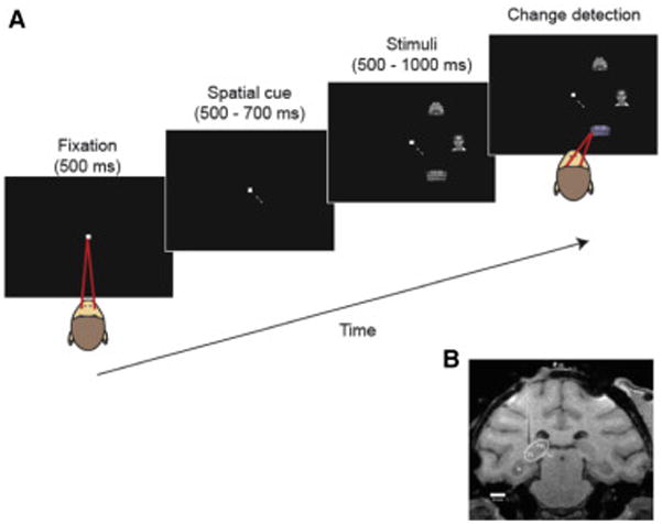
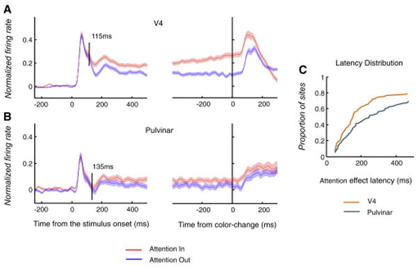
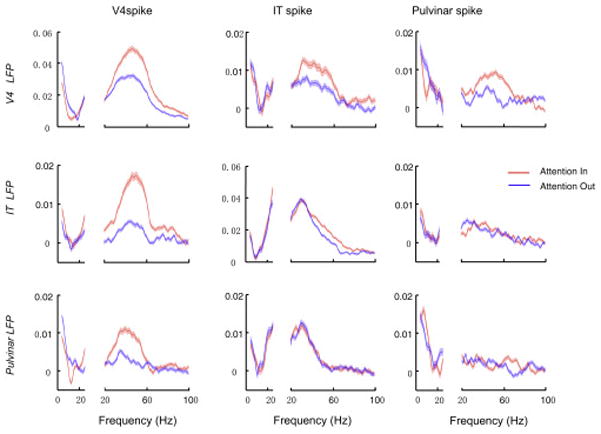
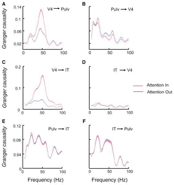
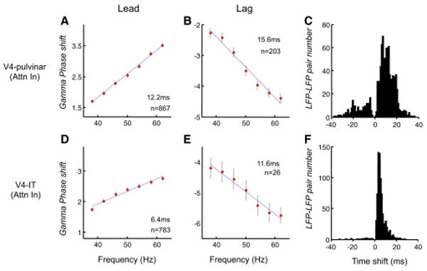
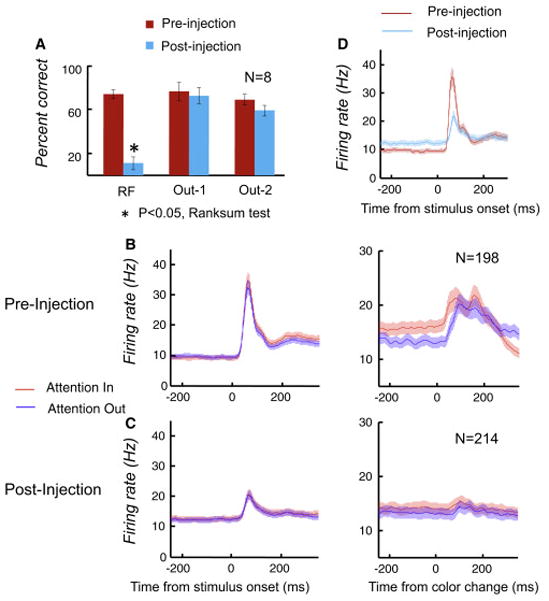
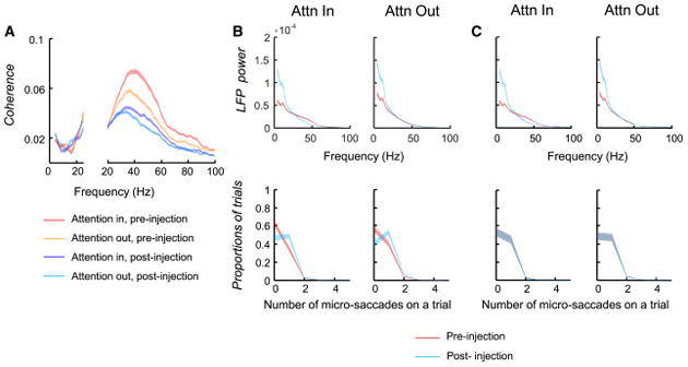
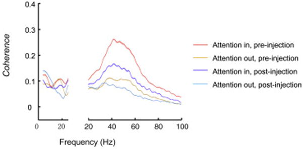
The ventro-lateral pulvinar is reciprocally connected with the visual areas of the ventral stream that are important for object recognition. To understand the mechanisms of attentive stimulus processing in this pulvinar-cortex loop, we investigated the interactions between the pulvinar, area V4, and IT cortex in a spatial-attention task. Sensory processing and the influence of attention in the pulvinar appeared to reflect its cortical inputs. However, pulvinar deactivation led to a reduction of attentional effects on firing rates and gamma synchrony in V4, a reduction of sensory-evoked responses and overall gamma coherence within V4, and severe behavioral deficits in the affected portion of the visual field. Conversely, pulvinar deactivation caused an increase in low-frequency cortical oscillations, often associated with inattention or sleep. Thus, cortical interactions with the ventro-lateral pulvinar are necessary for normal attention and sensory processing and for maintaining the cortex in an active state.


Figures

Figure 1
Figure 1
Task and recording sites. (a) Task in the cue-first condition. A spatial cue appeared that pointed to the upcoming target stimulus location, then 3 stimuli (target and 2 distracters) appeared on the contra-recording side of the screen. The monkey was rewarded for making a saccade to the target when it changed slightly in color, which occurred randomly from 500 to 1000ms after stimulus onset. (b) MRI image showing an electrode in place above the recording area in pulvinar.
Figure 2
Figure 2
Attentional modulation in V4 and pulvinar. (a) Normalized firing rates averaged across the V4 sites during ‘Attention In’ and ‘Attention Out’ conditions (n=152). Shading around average firing rates indicates the SEM (±). The black vertical line in the left plot marks the time when the Attention In and Attention Out responses reached a significant difference, and it was defined to be the attentional latency. (b) Population averages across pulvinar neurons (n=105). (c) The cumulative distribution of attentional latencies computed individually for V4 and pulvinar sites. The proportions of sites with attentional latencies <=50ms were combined.
Figure 3
Figure 3
Effects of attention on spike-LFP coherence. Population averages of the coherence in Attention In and Attention Out conditions were calculated between spike and LFP signals within and between V4, IT and pulvinar. The coherence at low frequencies (4–25 Hz) and higher frequencies was calculated and displayed separately. The SEM (±) of the population averages is indicated by the shading around the averages.
Figure 4
Figure 4
Granger causality between V4, pulvinar and IT LFPs in frequency domain. (a) V4 influence on pulvinar (Pulv). The SEM (±) of the population averages is marked by the shading around the averages. (b) Pulvinar influence on V4. (c) V4 influence on IT. (d) IT influence on V4. (e) Pulvinar influence on IT. (f) IT influence on pulvinar. Format in (b)–(f) is the same as the format in (a).
Figure 5
Figure 5
Directionality based on gamma frequency phase-shift across structures. (a) V4 led pulvinar in gamma phase. In 867 of 1608 V4 LFP pulvinar LFP pairs, the phase-lag of pulvinar to V4 increased linearly with the increasing frequency within the gamma range. The thick red dots represent the averaged phase-shifts across the 867 LFP pairs, and the SEM (±) of the averaged phase-shifts are marked by the vertical lines centered on these red dots. The regression line for these averaged phase-shifts is shown in blue, and the slope indicates that V4 led pulvinar by 12.2ms. (b) Pulvinar led V4 gamma phase. In 203 of 1608 V4 LFP – pulvinar LFP pairs, the phase-lag of pulvinar to V4 decreased linearly with the increasing frequency, and the slope indicated that pulvinar led V4 by 15.6ms. (c) Distribution of time shifts between V4 and pulvinar gamma oscillations. The positive time means that V4 gamma led pulvinar gamma. (d)-(e) show the phase-shift and the corresponding time-shift between V4 and IT gamma oscillations (totally 1072 pairs). The formats in (d),(e) and (f) are the same as the formats in (a), (b) and (c), respectively.
Figure 6
Figure 6
Effects of pulvinar deactivation on behavior and neuronal activity in V4. (a) Muscimol injection in pulvinar selectively lowered monkeys’ performance when the target was located in the RF of recorded V4 neurons, while performances at the other two locations outside of the RF (Out-1 and Out-2) were not affected. The averaged performance and SEM (±) of the 8 sessions are shown. (b) and (c) Population averages of firing rates during Attention In and Attention Out condition before and after muscimol injections(n=198 pre-injection; n=214 post-injection). During the last 250ms before the color-change, V4 neurons showed enhanced responses with attention before pulvinar injection (b), but this enhancement with attention was reduced after the injection (c). (d) Pulvinar deactivation reduced visual responses, but increased baseline activity before stimulus onset in V4. Attention Out responses before and after the injection are shown here.
Figure 7
Figure 7
Effects of pulvinar deactivation on coherence and LFP power in V4. (a) Population averages of V4 spike V4 LFP coherence in Attention In and Attention Out conditions before and after the muscimol injection (pre-injection, n=2177; post injection, n=2306). (b) Effects of the pulvinar deactivation on V4 LFP power without matching the number of microsaccades. The power spectrums were calculated in the last 256ms period prior to the color change. The upper plots show population averages of V4 LFP power spectrum before and after pulvinar deactivation. In both Attention In and Attention Out conditions, the low frequency LFP power after injection was significantly stronger than the power before injection (Wilcoxon signed rank test, P<0.05; n=153). The lower two plots show the distribution of proportions of trials with different number of microsaccades within the 256 ms period. There were slightly more microsaccades in the post-injection than in the pre-injection period. (c) Effects of the pulvinar deactivation on V4 LFP powers after matching the number of microsaccades. The effects on low frequency power were almost the same as those shown in (b).
Figure 8
Figure 8
Effects of pulvinar deactivation on coherence between V4 and IT. Population averages of V4 LFP – IT LFP coherence in Attention In and Attention Out conditions before and after the muscimol injection are shown (n=2048).

Comment in

  • Pondering the Pulvinar.
    Lakatos P, O'Connell MN, Barczak A. Lakatos P, et al. Neuron. 2016 Jan 6;89(1):5-7. doi: 10.1016/j.neuron.2015.12.022. Neuron. 2016. PMID: 26748085 Free PMC article.

References

    1. Arend I, Rafal R, Ward R. Spatial and temporal deficits are regionally dissociable in patients with pulvinar lesions. Brain. 2008;131:2140–2152. - PubMed
    1. Baizer JS, Desimone R, Ungerleider LG. Comparison of subcortical connections of inferior temporal and posterior parietal cortex in monkeys. Vis Neurosci. 1993;10:59–72. - PubMed
    1. Baldauf D, Desimone R. Neural mechanisms of object-based attention. Science. 2014;344:424–427. - PubMed
    1. Bastos AM, Vezoli J, Fries P. Communication through coherence with inter-areal delays. Curr Opin Neurobiol. 2014;31C:173–180. - PubMed
    1. Bender DB, Butter CM. Comparison of the effects of superior colliculus and pulvinar lesions on visual search and tachistoscopic pattern discrimination in monkeys. Exp Brain Res. 1987;69:140–154. - PubMed

Publication types