

Oral manifestation of tuberculosis: a case-report
- PMID: 26748230
- PMCID: PMC9427557
- DOI: 10.1016/j.bjid.2015.12.001
Oral manifestation of tuberculosis: a case-report
Abstract
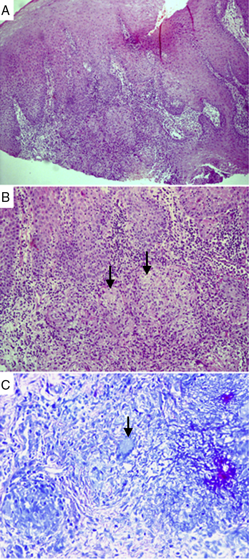
The present case-report describes tuberculosis on the oral mucosa, in a rare manifestation of the disease. The importance of appropriate diagnosis and awareness of the clinical manifestations is highlighted. Oral lesions seem to occur as chronic ulcers, nodular or granular areas, and rare, firm leukoplakia regions. Most extra-pulmonary lesions represent secondary infections of a primary lung infectious focus; therefore, early and accurate diagnosis is required for planning of the best treatment and strategies to control the disease.
Keywords: Mycobacterium infection; Mycobacterium tuberculosis; Oral lesion; Oral tuberculosis.
Copyright © 2016. Published by Elsevier Editora Ltda.
Figures

Similar articles
-
[Oral manifestation of miliary tuberculosis].Mund Kiefer Gesichtschir. 2005 May;9(3):180-3. doi: 10.1007/s10006-005-0596-6. Mund Kiefer Gesichtschir. 2005. PMID: 15726436 German.
-
Lupus vulgaris of the oral mucosa. Report of 4 cases associated with asymptomatic pulmonary tuberculosis.Dermatologica. 1981;162(3):183-90. Dermatologica. 1981. PMID: 7250462
-
Oral mucosal ulceration: a manifestation of previously undiagnosed pulmonary tuberculosis.J Am Dent Assoc. 2004 Mar;135(3):336-40. doi: 10.14219/jada.archive.2004.0184. J Am Dent Assoc. 2004. PMID: 15058623
-
Primary tuberculosis of the oral cavity in an elderly nonimmunosuppressed patient: case report and review of the literature.Arch Otolaryngol Head Neck Surg. 2008 Oct;134(10):1107-9. doi: 10.1001/archotol.134.10.1107. Arch Otolaryngol Head Neck Surg. 2008. PMID: 18936360 Review. No abstract available.
-
Tuberculosis of the oral cavity: a case report.Quintessence Int. 1997 Nov;28(11):745-7. Quintessence Int. 1997. PMID: 9573865 Review.
Cited by
-
Primary Tuberculosis of Buccal and Labial Mucosa: Literature Review and a Rare Case Report of a Public Health Menace.Case Rep Dent. 2023 Oct 5;2023:6543595. doi: 10.1155/2023/6543595. eCollection 2023. Case Rep Dent. 2023. PMID: 37842328 Free PMC article.
-
A buccal mucosa ulcer as the first sign of tuberculosis.J Oral Maxillofac Pathol. 2022 Jul-Sep;26(3):399-403. doi: 10.4103/jomfp.jomfp_443_21. Epub 2022 Oct 17. J Oral Maxillofac Pathol. 2022. PMID: 36588851 Free PMC article.
-
Clinical study of tuberculosis in the head and neck region-11 years' experience and a review of the literature.Emerg Microbes Infect. 2018 Jan 10;7(1):4. doi: 10.1038/s41426-017-0008-7. Emerg Microbes Infect. 2018. PMID: 29323108 Free PMC article. Review.
-
Orofacial Bacterial Infectious Diseases: An Update.J Int Soc Prev Community Dent. 2017 Oct;7(Suppl 2):S61-S67. doi: 10.4103/jispcd.JISPCD_290_17. Epub 2017 Oct 30. J Int Soc Prev Community Dent. 2017. PMID: 29184830 Free PMC article. Review.
-
A case of oral tuberculous ulcer and literature review.Clin Case Rep. 2023 Dec 5;11(12):e8216. doi: 10.1002/ccr3.8216. eCollection 2023 Dec. Clin Case Rep. 2023. PMID: 38076017 Free PMC article.
References
-
- Santiago R.A., Gueiros L.A., Porter S.R., et al. Prevalence of oral lesions in Brazilian patients with tuberculosis. Indian J Dent Res. 2013;24:245–248. - PubMed
-
- Kakisi O.K., Kechagia A.S., Kakisis I.K., et al. Tuberculosis of the oral cavity: a systematic review. Eur J Oral Sci. 2010;118:103–109. - PubMed
-
- Vaid S., Lee Y.Y., Rawat S., et al. Tuberculosis in the head and neck: a forgotten differential diagnosis. Clin Radiol. 2010;65:73–81. - PubMed
-
- González Martín J., García-García J.M., Anibarro L., et al. Consensus document on the diagnosis, treatment and prevention of tuberculosis. Arch Bronconeumol. 2010;46:255–274. - PubMed
Publication types
MeSH terms
LinkOut - more resources
Full Text Sources
Other Literature Sources
