Review
. 2016 Feb:91:1-5.
doi: 10.1016/j.yjmcc.2015.12.031. Epub 2015 Dec 31.

Origins of cardiac fibroblasts

Affiliations
Review

Origins of cardiac fibroblasts

Thomas Moore-Morris et al. J Mol Cell Cardiol. 2016 Feb.

Abstract

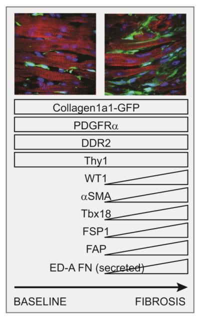
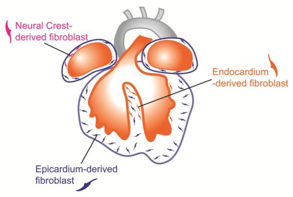
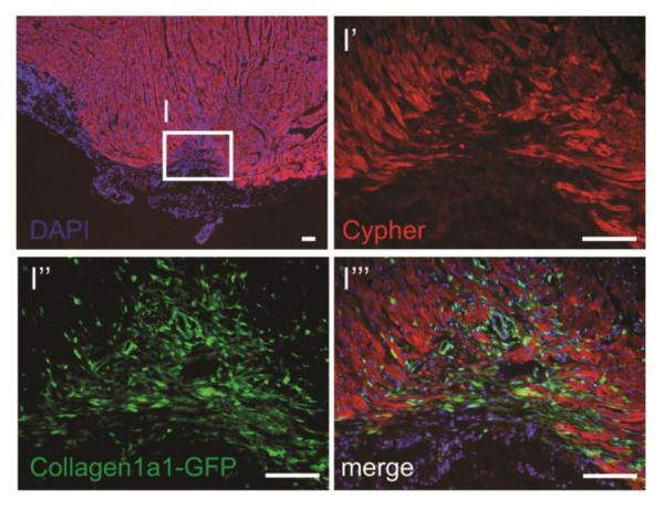
Cardiac fibroblasts produce the extracellular matrix (ECM) scaffold within which the various cellular components of the heart are organized. As well as providing structural support, it is becoming evident that the quality and quantity of ECM is a key factor for determining cardiac cell behavior during development and in pathological contexts such as heart failure involving fibrosis. Cardiac fibroblasts have long remained a poorly characterized cardiac lineage. Well characterized markers are now paving the way for a better understanding of the roles of these cells in various developmental and disease contexts. Notably, the relevance of processes including endothelial-tomesenchymal transition and the recruitment of circulating fibroblast progenitors in heart failure has been challenged. This review describes the latest findings on cardiac fibroblast markers and developmental origins, and discusses their importance in myocardial remodeling. Effective modulation of cardiac fibroblast activity would likely contribute to successful treatment of various cardiac disorders.

Keywords: Cardiac lineage; Fibroblast; Fibroblast markers; Fibrosis; Hypertrophy; Myocardial infarction.


Figures

Figure 1
Figure 1
Markers expressed by fibroblasts prior to and during fibrosis. Collagen1a1-GFP and PDGFRα are constitutively expressed by fibroblasts in normal and pathological contexts. Subsets of fibroblasts up-regulate a number of additional markers in fibrotic conditions, including αSMA and WT1, whose expression depends on the disease context and the location, notably with respect to large coronary vessels. Images: Cypher (myocyte markers, red), Collagen1a1-GFP(green), DAPI (blue).
Figure 2
Figure 2
Fibroblasts from cardiac lineages populate the heart during embryonic development. Most fibroblasts derived from the epicardium (blue), a protective epithelial layer that entirely covers the four cardiac chambers. Smaller populations are derived from the endocardium (orange) and cardiac neural crest, and are mostly found in the interventricular septum and right atrium, respectively.
Figure 3
Figure 3
Collagen type I expressing fibroblasts (green) are active in the area of regeneration, tightly associated with myocytes (Cypher+, red) following resection of neonatal mouse heart (unpublished observations, Evans S.M. et al.). Images represent the apex of a heart 2 weeks after resection, during the window of regeneration previously described [2]. Scale bars represent 100μm.

References

    1. Roger VL, Go AS, Lloyd-Jones DM, Benjamin EJ, Berry JD, Borden WB, et al. Heart disease and stroke statistics--2012 update: a report from the American Heart Association. Circulation. 2012;125:e2–e220. - PMC - PubMed
    1. Porrello ER, Mahmoud AI, Simpson E, Hill JA, Richardson JA, Olson EN, et al. Transient regenerative potential of the neonatal mouse heart. Science (New York, NY) 2011;331:1078–80. - PMC - PubMed
    1. Moore-Morris T, Guimaraes-Camboa N, Banerjee I, Zambon AC, Kisseleva T, Velayoudon A, et al. Resident fibroblast lineages mediate pressure overload-induced cardiac fibrosis. The Journal of clinical investigation. 2014;124:2921–34. - PMC - PubMed
    1. Ali SR, Ranjbarvaziri S, Talkhabi M, Zhao P, Subat A, Hojjat A, et al. Developmental Heterogeneity of Cardiac Fibroblasts Does Not Predict Pathological Proliferation and Activation. Circulation research. 2014 - PubMed
    1. Ieda M, Tsuchihashi T, Ivey KN, Ross RS, Hong TT, Shaw RM, et al. Cardiac fibroblasts regulate myocardial proliferation through beta1 integrin signaling. Developmental cell. 2009;16:233–44. - PMC - PubMed

Publication types

MeSH terms