. 2017 Jan;27(1):26-35.
doi: 10.1111/bpa.12347. Epub 2016 Feb 22.

Mild Malformation of Cortical Development with Oligodendroglial Hyperplasia in Frontal Lobe Epilepsy: A New Clinico-Pathological Entity

Affiliations

Mild Malformation of Cortical Development with Oligodendroglial Hyperplasia in Frontal Lobe Epilepsy: A New Clinico-Pathological Entity

Johannes Schurr et al. Brain Pathol. 2017 Jan.

Abstract

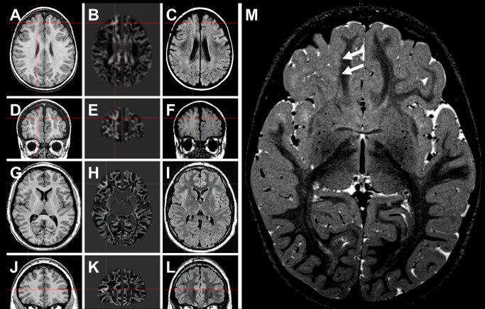
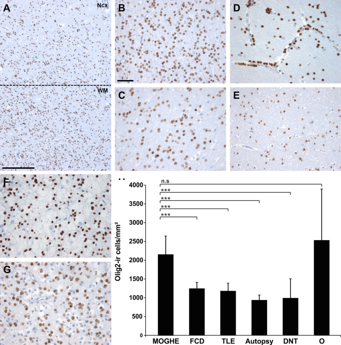
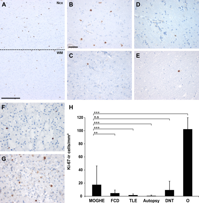
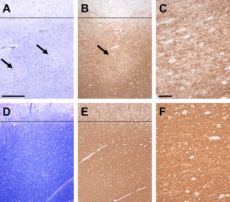
The histopathological spectrum of human epileptogenic brain lesions is widespread including common and rare variants of cortical malformations. However, 2-26% of epilepsy surgery specimens are histopathologically classified as nonlesional. We hypothesized that these specimens include also new diagnostic entities, in particular when presurgical magnetic resonance imaging (MRI) can identify abnormal signal intensities within the anatomical region of seizure onset. In our series of 1381 en bloc resected epilepsy surgery brain specimens, 52 cases could not be histopathologically classified and were considered nonlesional (3.7%). An increase of Olig2-, and PDGFR-alpha-immunoreactive oligodendroglia was observed in white matter and deep cortical layers in 22 of these patients (42%). Increased proliferation activity as well as heterotopic neurons in white matter were additional histopathological hallmarks. All patients suffered from frontal lobe epilepsy (FLE) with a median age of epilepsy onset at 4 years and 16 years at epilepsy surgery. Presurgical MRI suggested focal cortical dysplasia (FCD) in all patients. We suggest to classify this characteristic histopathology pattern as "mild malformation of cortical development with oligodendroglial hyperplasia (MOGHE)." Further insights into pathomechanisms of MOGHE may help to bridge the diagnostic gap in children and young adults with difficult-to-treat FLE.

Keywords: epilepsy; frontal lobe; hyperplasia; neuropathology; oligodendrocytes.


Figures

Figure 1
Figure 1
Neuroimaging findings in MOGHE. A–F. Patient #2 (Table 1). G–L. Patient #13 (Table 1). In the latter patient, the lesion became evident only after morphometric MRI analysis (junction image in 2nd column). 23 Red crosses highlight a suspected lesion in T1 (1st column), and postprocessed junction maps (2nd column). 3rd column: FLAIR sequences at same section planes. M. Three‐year old female patient with band‐like signal alteration at the gray‐white matter junction in right frontal lobe (arrows in T2 weighted turbo echo spin image) and histopathologically confirmed MOGHE.
Figure 2
Figure 2
Oligodendroglial cell densities in epilepsy specimens and controls. A. Olig2 immunohistochemistry at the gray (NCx) ‐white matter (WM) junction in MOGHE (dotted line). Scale bar = 500 μm. B–G. higher magnification from white matter regions in different pathology samples and post‐mortem controls (autopsy; E). B. MOGHE; (C) FCD ILAE Type I, (D) temporal lobe epilepsy (TLE) with perivascular clustering of oligodendroglia‐like cells 25, 39; (F) DNT (WHO I°), (G) oligodendroglioma (O; WHO II°). Scale bar in B = 20 μm, applies also to C–G. H. a statistically significant increase was detected for Olig2‐positive cells in MOGHE and oligodendrogliomas (O). *** = P < 0.001, n.s. = not significant.
Figure 3
Figure 3
Proliferation activity in epilepsy specimens and controls. A. Ki‐67 immunohistochemistry at gray (NCx) ‐ white matter (WM) junction (dotted line) in MOGHE. Scale bar = 500 μm. B–G. higher magnification from white matter regions in different pathology samples and post‐mortem controls (autopsy; E). B. MOGHE; C. FCD ILAE Type I, D. temporal lobe epilepsy (TLE) with perivascular clustering of oligodendroglia‐like cells; F. DNT (WHO I°), G. oligodendroglioma (WHO II°). Scale bar in B = 20 μm, applies also to C–G. H. A statistically significant increase of Ki‐67 positive cells was detected in MOGHE compared with FCD I, TLE and autopsy controls (** = P < 0.01; *** = P < 0.001). A similar proliferation activity was encountered in DNTs (WHO I°, n.s. = not significant), whereas oligodendrogliomas (O) showed significantly higher proliferation (P < 0.001).
Figure 4
Figure 4
Decreased myelin staining densities in MOGHE. A. Nissl‐LFB staining in MOGHE compared with nonepileptic controls (D) revealed decreased staining intensities in juxtacortical areas (arrows). Dotted lines indicate gray‐white matter boundary. B. CNPase immunoreactivity was also decreased, often occurring with a pachty pattern (arrow). C. higher magnification of area indicated by arrow in B. E/F. a homogeneous distribution of CNPase immunoreactivity was observed in our control series. Scale bar in A: 400 μm, applies also for B,E; Scale bar in C: 50 μm, applies also for F.
Figure 5
Figure 5
Clinico‐pathological correlation in MOGHE. A. increased oligodendroglial cell densities did not change significantly with duration of epilepsy (Spearman rank test, not significant n.s.). B. significantly higher proliferation activity of Olig2‐positive cells was identified at younger age of operation (Spearman rank test, ** P < 0.01).

Similar articles

Cited by

References

    1. Armstrong DD (1993) The neuropathology of temporal lobe epilepsy. J Neuropathol Exp Neurol 52:433–443. - PubMed
    1. Aronica E, Sandau US, Iyer A, Boison D (2013) Glial adenosine kinase ‐ a neuropathological marker of the epileptic brain. Neurochem Int 63:688–695. - PMC - PubMed
    1. Barkovich AJ, Guerrini R, Kuzniecky RI, Jackson GD, Dobyns WB (2012) A developmental and genetic classification for malformations of cortical development: update 2012. Brain 135(Pt 5):1348–1369. - PMC - PubMed
    1. Bien CG, Raabe AL, Schramm J, Becker A, Urbach H, Elger CE (2013) Trends in presurgical evaluation and surgical treatment of epilepsy at one centre from 1988‐2009. J Neurol Neurosurg Psychiatry 84:54–61. - PubMed
    1. Blumcke I, Aronica E, Urbach H, Alexopoulos A, Gonzalez‐Martinez JA (2014) A neuropathology‐based approach to epilepsy surgery in brain tumors and proposal for a new terminology use for long‐term epilepsy‐associated brain tumors. Acta Neuropathol 128:39–54. - PMC - PubMed

Publication types

MeSH terms

Substances