

Review
doi: 10.1016/j.stem.2015.12.012.
Material Cues as Potent Regulators of Epigenetics and Stem Cell Function
Affiliations
- PMID: 26748755
- PMCID: PMC5409508
- DOI: 10.1016/j.stem.2015.12.012
Item in Clipboard
Review
Material Cues as Potent Regulators of Epigenetics and Stem Cell Function
Cell Stem Cell.
.
Abstract
Biophysical signals act as potent regulators of stem cell function, lineage commitment, and epigenetic status. In recent years, synthetic biomaterials have been used to study a wide range of outside-in signaling events, and it is now well appreciated that material cues modulate the epigenome. Here, we review the role of extracellular signals in guiding stem cell behavior via epigenetic regulation, and we stress the role of physicochemical material properties as an often-overlooked modulator of intracellular signaling. We also highlight promising new research tools for ongoing interrogation of the stem cell-material interface.
Copyright © 2016 Elsevier Inc. All rights reserved.
Figures
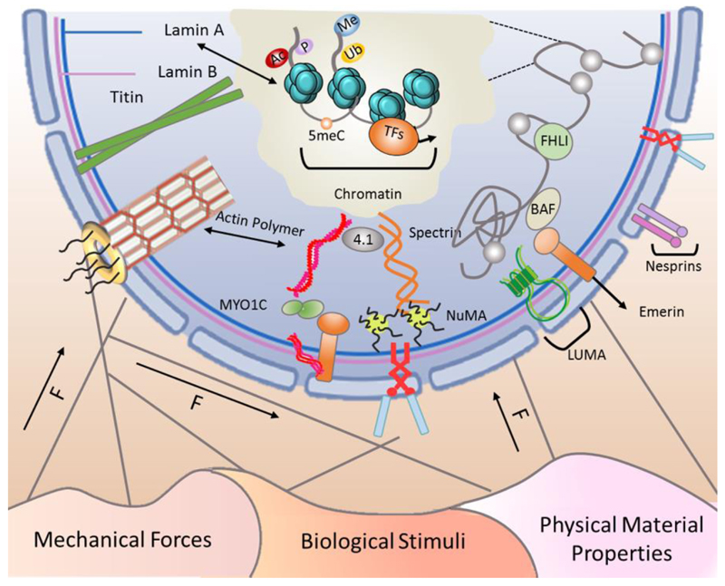
The cell is an interconnected entity with cytoskeletal components linking the membrane to the nucleus. Cells sense mechanical forces in their environment and propagate the forces along the cytoskeleton to the nucleus in order to alter epigenetic status and gene expression profile in response to different biophysical stimuli. Here, we classify these cues as mechanical forces, biological stimuli, and physical material properties. The nuclear envelope contains components of the LINC (Linker of Nucleoskeleton and Cytoskeleton) complex including nesprin and SUN proteins, emerin, and LUMA. These LINC complex components act as receivers and transmitters of mechanical forces to the chromatin and the nucleoskeleton, which includes polymerized actin, nuclear mitotic apparatus protein (NuMA), intermediate filaments, spectrins, protein 4.1, titin, A- and B-type lamins, and nuclear pore complex (NPC)-linked filaments. External forces induce mechanosensitive changes in the nucleoskeletal complexes that, in turn, alter the epigenome and chromatin accessibility. Therefore, these extracellular signals are perceived at the nuclear level as the cell adapts its transcriptome in response to the signals it perceives.
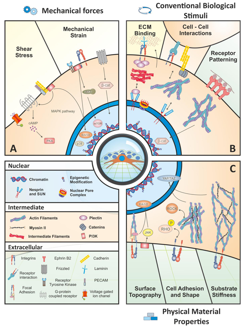
Extrinsic cues can be divided into three general categories: (A) Direct application of mechanical forces, such as shear stress and mechanical strain are relatively well-characterized examples of how biophysical signals from the microenvironment activate specific signaling pathways and alter the epigenetic status. Shear stress is transduced through multiple mechanisms, and a few key examples listed here include voltage-gated ion channels, G protein-coupled receptors (GPCRs), and cell adhesion molecules such as cadherins and platelet-endothelial cell adhesion molecule (PECAM). These sensors activate classical elements such as the MAPK pathway, which leads to the nuclear translocation of ERK1/2 and p38 to act on downstream target genes, and activation of PI3K and PKA. Mechanical strain sensing involves a myriad of receptor tyrosine kinases as well as the Wnt receptor, Frizzled, and an array of integrins. These extracellular mediators can activate the PI3K/AKT pathway, which stimulates mTOR activity. (B) Conventional biological stimuli include cell-ECM and cell-cell interactions, and receptor patterning/clustering. These events exhibit a feedback response: the cell receives specific input from one of these effectors, and in turn changes the signal by altering the composition or organization of the ECM or responds with an additional signal to a neighboring cell and propagating the message into something dynamic. Cell-ECM interactions are dominated by integrins at the cell surface that exert their effect directly on the nucleus via cytoskeletal filaments. Cell-cell interactions act in a similar manner but also include cell adhesion molecules such as cadherins. Receptor patterning occurs naturally in the sense that the spatial distribution of signaling or adhesion molecules affects cell behavior. In a synthetic system, receptors such as Ephrin-B2 can be encouraged to cluster by presenting the ligand at varying density along a physically-linked backbone, which stimulates a stronger response than a single ligand alone; in this case, Ephrin-B2 stimulates the stabilization and nuclear translocation of β-catenin. (C) Physical material properties at the macro, micro and nano scales include surface topography, cell adhesion and shape, and substrate stiffness. Topography of the material surface, such as the distribution and regularity of nanopit spacing, activates signaling pathways such as JNK and Erk1/2 and affects cytoskeletal tension. Cell adhesion and shape can be controlled by altering the adhesive sites on a synthetic substrate. This effect regulates cell fate decisions in hMSCs through the Rho/ROCK pathway, which is necessary for the activity of the YAP and TAZ transcription factors. Finally, the physical stiffness of the extracellular substrate heavily influences cell behaviors, such as fate decisions, and this signal is also transduced through YAP/TAZ and the actin cytoskeleton.
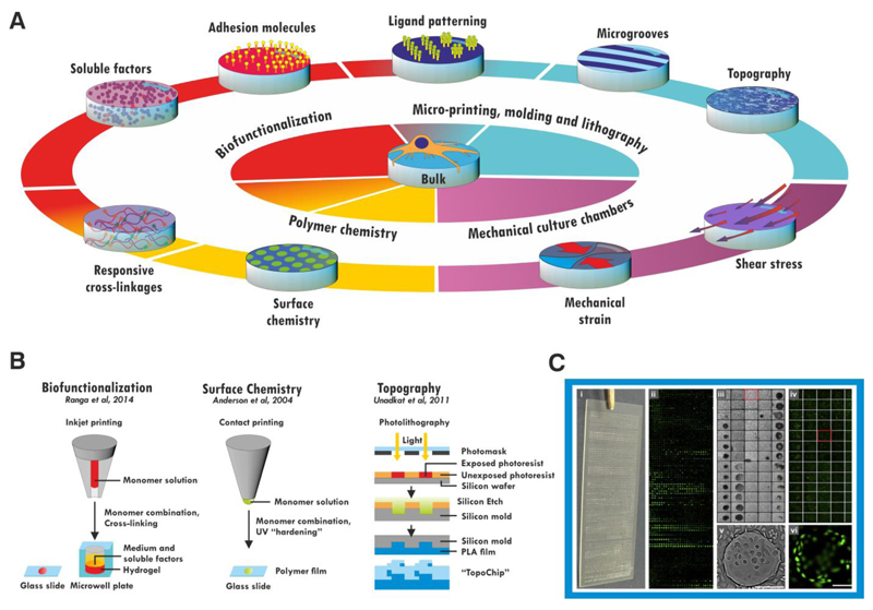
A bulk material can be further functionalized by a number of routes (A). Soluble biomolecules can be incorporated into the bulk of the material or presented on the surface, and hydrogels may be made biologically responsive by use of protease-degradable crosslinkages. Micropatterning techniques allow these cues to be patterned within the material, and also allow the material itself to be patterned into grooves or more complex topographical cues on the micro- or nano-scales. The chemistry of the material itself can be altered (e.g. by blending with other polymers) to confer responsiveness or alter the cell-material surface interface. Materials may also be used to subject the cells on them to mechanical strain or shear stress in combination with appropriate culture chambers. These modifications can be made in combination to generate combinatorial materials. Some of these cues have been screened in high-throughput studies (B). Surface chemistry can be investigated by microcontact printing and UV crosslinking of monomer combinations on a glass slide. Biofunctionalizations have been screened using micro-inkjet systems to generate hydrogels within a 1536-well plate or on a glass slide. Topographical cues have been investigated using photolithography to etch microscale geometric cues onto a poly-lactic acid (PLA) chip. The response of cells is screened by imaging (C) the entire surface, generating values for cell attachment or proliferation (iii, v) and fluorescent reporter expression (ii, iv, vi) for each individual material spot. Figure C adapted with permission from (Zhang et al., 2009), scale bar: 200 μm.
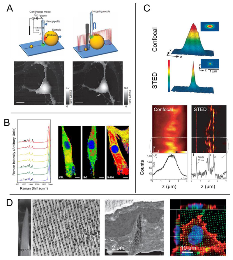
(A) Scanning ion conductance microscopy (SICM) uses a nanopipette to produce an image of a living cell without physical interaction. Two different modes of SICM are shown here: continuous mode in which the probe does not significantly change its z-position, and hopping mode in which the probe is constantly approaching the sample and then retreating. In hopping mode, small features that are not observed with continuous mode can become spatially resolved (Scale bars, 10 μm). (B) Raman spectroscopy produces a chemical ‘fingerprint’ of the cell(s) or tissue that is studied, providing information about the specific chemical bonds present in the sample. This information can then be used to produce a 2D or 3D map that shows the localization of certain classes of molecules. For example, bioactive glass materials with a significant amount of strontium ion (Sr100) lead to a shift in the lipid distribution in MSCs (lipid, red) (Scale bars, 10 μm). (C) Stimulated emission depletion (STED) microscopy overcomes the diffraction limit typically associated with light-based imaging techniques. The focal point of a STED microscope is much smaller (bottom graph) than that of a standard confocal (top graph). This allows for enhanced spatial resolution, such as imaging the microtubule network in monolayer cell culture. Typical confocal (left image) produces an image in which the network cannot be observed, but sub-diffraction STED imaging allows for crisp visualization of the subcellular processes. (D) High-aspect ratio nanoneedles can be produced into high density, 2D arrays to act as cell culture substrates. HeLa cells cultured on nanoneedles interact directly with the underlying material, and the needles physically deform both the cell membrane as well as the nucleus. Figures adapted with permission from: (A) Nature Publishing Group (NPG) Ref: (Novak et al., 2009); (B) Ref: (Autefage et al., 2015); (C) NPG Ref: (Dyba et al., 2003); (D) Reprinted with permission from Ref: (Chiappini et al., 2015c). Copyright 2015 American Chemical Society; NPG Ref: (Chiappini et al., 2015b), and Ref: (Chiappini et al., 2015a).
References
-
- Alabert C, Bukowski-Wills JC, Lee SB, Kustatscher G, Nakamura K, de Lima Alves F, Menard P, Mejlvang J, Rappsilber J, Groth A. Nascent chromatin capture proteomics determines chromatin dynamics during DNA replication and identifies unknown fork components. Nat Cell Biol. 2014;16:281–293. - PMC - PubMed
-
- Anderson DG, Levenberg S, Langer R. Nanoliter-scale synthesis of arrayed biomaterials and application to human embryonic stem cells. Nat Biotechnol. 2004;22:863–866. - PubMed
Publication types
MeSH terms
Substances
Grants and funding
LinkOut - more resources
Full Text Sources
Other Literature Sources
Medical
