Review
. 2016 Mar 15;594(6):1529-51.
doi: 10.1113/JP271480. Epub 2016 Feb 19.

Proton detection and breathing regulation by the retrotrapezoid nucleus

Affiliations
Review

Proton detection and breathing regulation by the retrotrapezoid nucleus

Patrice G Guyenet et al. J Physiol. .

Abstract

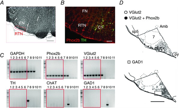
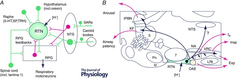
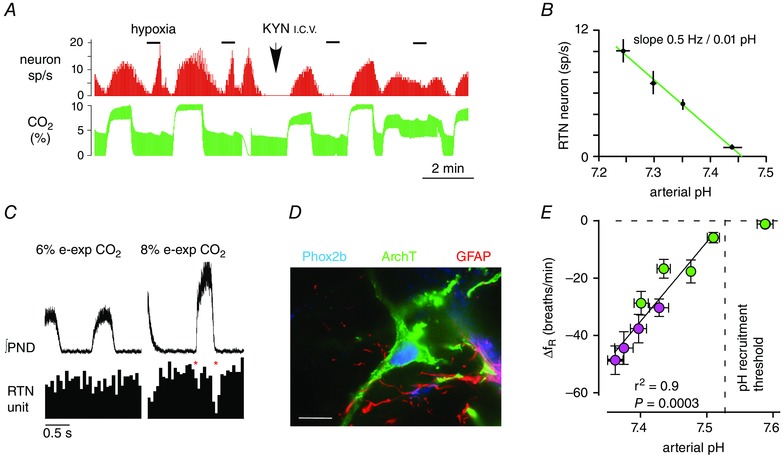
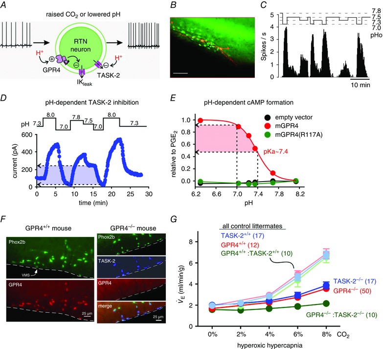
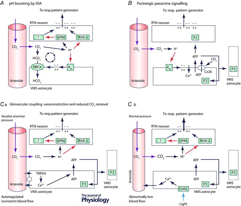
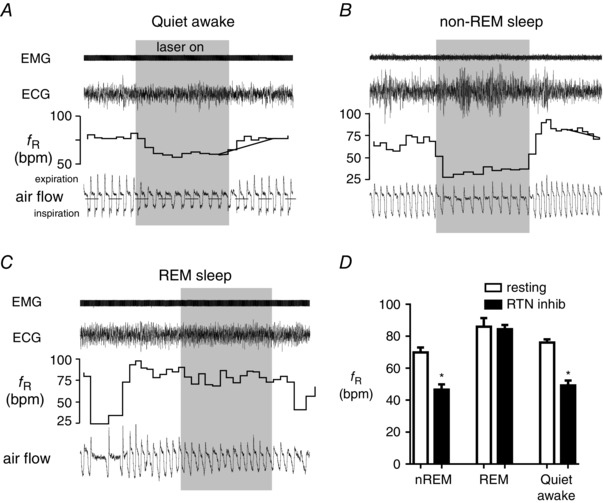
We discuss recent evidence which suggests that the principal central respiratory chemoreceptors are located within the retrotrapezoid nucleus (RTN) and that RTN neurons are directly sensitive to [H(+) ]. RTN neurons are glutamatergic. In vitro, their activation by [H(+) ] requires expression of a proton-activated G protein-coupled receptor (GPR4) and a proton-modulated potassium channel (TASK-2) whose transcripts are undetectable in astrocytes and the rest of the lower brainstem respiratory network. The pH response of RTN neurons is modulated by surrounding astrocytes but genetic deletion of RTN neurons or deletion of both GPR4 and TASK-2 virtually eliminates the central respiratory chemoreflex. Thus, although this reflex is regulated by innumerable brain pathways, it seems to operate predominantly by modulating the discharge rate of RTN neurons, and the activation of RTN neurons by hypercapnia may ultimately derive from their intrinsic pH sensitivity. RTN neurons increase lung ventilation by stimulating multiple aspects of breathing simultaneously. They stimulate breathing about equally during quiet wake and non-rapid eye movement (REM) sleep, and to a lesser degree during REM sleep. The activity of RTN neurons is regulated by inhibitory feedback and by excitatory inputs, notably from the carotid bodies. The latter input operates during normo- or hypercapnia but fails to activate RTN neurons under hypocapnic conditions. RTN inhibition probably limits the degree of hyperventilation produced by hypocapnic hypoxia. RTN neurons are also activated by inputs from serotonergic neurons and hypothalamic neurons. The absence of RTN neurons probably underlies the sleep apnoea and lack of chemoreflex that characterize congenital central hypoventilation syndrome.


Figures

Figure 1
Figure 1. Location and phenotype of RTN neurons
A, transverse section through the caudal end of the facial motor nucleus (FN, Sprague–Dawley adult rat; bregma −11.6 mm; myelin stain; scale 500 μm). Box showing approximate location of retrotrapezoid nucleus. B, RTN identified as Phox2b‐immunoreactive (Phox2b‐ir), non‐catecholaminergic (TH‐negative) neurons in a transverse section of medulla oblongata (bregma −11.6 mm; adult Sprague–Dawley rat; scale bar: 100 μm). The C1 (adrenergic/glutamatergic) neurons also express Phox2b (reproduced from Guyenet, 2008). C, single‐cell RT‐PCR data showing presence of Phox2b and VGlut2 transcripts in enhanced green fluorescent protein (eGFP)‐expressing neurons dissociated from the RTN of a Phox2b‐eGFP mouse (lanes 1–6), including three pH‐sensitive neurons examined after recording (lanes 4–6); the RTN neurons did not express tyrosine hydroxylase (TH), choline acetyl‐transferase (ChAT), or glutamate decarboxylase 1 (GAD1). Control experiments verified detection of the following: TH (with Phox2b and VGlut2) in a C1 neuron (lane 7); ChAT (and Phox2b) in a facial motoneuron (lane 8); and GAD1 in a striatal medium spiny cell (lane 9). Glyceraldehyde‐3‐phosphate dehydrogenase (GAPDH) expression was seen in all cells, and negative controls for each PCR reaction included bath solution (lane 10) and water (lane 11) substituted for cell contents (reproduced from Wang et al. 2013 b). D, plots of glutamatergic (VGlut2‐mRNA+) and GABAergic (GAD1‐mRNA+) neurons in a representative transverse section through the rat's medulla oblongata (bregma level −11.4 mm; calibration 500 μm). Box shows predominance of glutamatergic neurons in RTN. All Phox2b‐ir neurons express VGlut2 (filled circles); none contain GAD1. The Phox2b+ neurons, most of which are non‐catecholaminergic at this coronal level, are closely surrounded by Phox2b‐negative glutamatergic neurons and Phox2b‐negative GABAergic neurons (reproduced from Stornetta et al. 2006). Amb, nucleus ambiguus, compact part; sp5, spinal trigeminal tract; 7, facial motor nucleus; py, pyramidal tract.
Figure 2
Figure 2. Inputs and outputs of RTN neurons
A, main connections of RTN neurons (green, excitatory; magenta inhibitory). The RPG includes the ventral respiratory column (VRC) and the dorsolateral pons. The monosynaptic nature of the RTN inputs shown in this figure has not yet been confirmed by ultrastructural evidence. B, parasagittal section through the rat's brain (∼1.8 mm lateral to the midline) illustrating the location of RTN neurons, their axonal projections and the putative function of each projection. Abbreviations: 5‐HT, serotonin; Exp, active (abdominal) expiration; f R, breathing frequency; Insp, inspiration; KF, Kölliker–Fuse nucleus; lPBN, lateral parabrachial nuclei; LRt, lateral reticular nucleus; NA, nucleus ambiguus; NTS, nucleus of the solitary tract; OAE, oscillator for active expiration; Pn, pontine nuclei; RPG, respiratory pattern generator; SARs, stretch‐activated receptors; SP, substance P; TRH, thyrotropin‐releasing hormone; tz, trapezoid body; VRC, ventral respiratory column (incl. its four subdivisions, from rostral to caudal: Bötzinger, preBötzinger complex, rVRG and cVRG).
Figure 3
Figure 3. RTN neurons encode PCO2 in vivo
A, single RTN neuron recorded in a halothane‐anaesthetized vagotomized rat. The neuron is activated by elevated CO2 (hypercapnia) and by hypoxia (sp/s, spikes s−1). The effect of hypoxia is selectively eliminated by administration of kynurenic acid (KYN, i.c.v.) to block glutamate transmission in the brainstem (reproduced from Mulkey et al. 2004). B, relationship between the discharge rate of RTN neurons (n = 12) and arterial pH in halothane‐anaesthetized rats in which glutamatergic transmission was impaired (by KYN, i.c.v.). C, respiratory modulation of an RTN neuron in a vagotomized halothane‐anaesthetized rat. The respiratory modulation was enhanced by hypercapnia (right panel). This particular neuron exhibits two periods of reduced firing probability per breathing cycle, one at the onset of inspiration, the other during post‐inspiration (asterisks). Top trace, averaged rectified phrenic nerve discharge (∫PND; 100 sweeps triggered on PND upstroke). Bottom trace: event‐triggered histogram of neuron action potentials (50 ms bins) (redrawn from Guyenet et al. 2005). D, example of one RTN neuron transduced with archaerhodopsin from Halorubrum strain TP009, version 3.0 (ArchT3.0) (astrocytes, identified by the presence of glial fibrillary acid protein, GFAP, do not express the opsin). E, the reduction of breathing frequency caused by bilateral opto‐inhibition of RTN neurons plotted as a function of arterial pH. Bilateral inhibition of archaerhodopsin (ArchT3.0)‐transduced RTN neurons decreases breathing frequency in proportion to plasma pH in conscious rats. Plasma pH was manipulated by lowering or increasing FIO2 (green symbols) or by combining various degrees of respiratory alkalosis on a background of metabolic acidosis by administering acetazolamide (pink symbols). Adapted from Basting et al. (2015).
Figure 4
Figure 4. RTN neurons directly encode [H+]
A, molecular basis of the intrinsic proton sensitivity of RTN neurons (reproduced from Kumar et al. 2015). B, RTN neurons identified in a Phox2b‐eGFP transgenic mouse (green cells). Two RTN neurons were filled with biotinamide after intracellular recording (orange; scale bar: 50 μm). C, right, example of pH‐dependent firing rate of one RTN neuron recorded in a Phox2b‐eGFP mouse brain slice (reproduced from Lazarenko et al. 2009). D, current elicited in a HEK293 cell transiently transfected with TASK‐2. Note the large inward shift in holding current elicited by changing extracellular pH from neutral (7.5) to acid (7.0). E, representative pH‐dependent accumulation of cAMP in HEK293 cells transiently transfected with mGPR4 (red curve), by comparison with cell transfected with empty vector (black) or mGPR4(R117A), a signalling‐deficient receptor mutant (green). The response is normalized to cAMP accumulation elicited in these cells by a saturating concentration of PGE2, which activates an endogenous receptor. F, left two panels show colocalization of Phox2b (top) and GPR4 transcripts (bottom) in RTN neurons in a wild‐type mouse. The four panels on the right show an experiment performed in a GPR4 knockout mouse. From top to bottom: Phox2b mRNA, TASK‐2 mRNA, absence of GPR4 transcripts and merged image showing colocalization of Phox2b and TASK‐2 transcripts. VMS, ventral medullary surface. G, central respiratory chemoreflex (V˙E, minute ventilation; breathing stimulation elicited by hyperoxic hypercapnia) is reduced by 65% in TASK‐2 and in GPR4 knockout mice relative to control littermates (n provided in parentheses). GPR4−/−:TASK‐2−/− mice deleted for both genes show 90% reduction (with sample size increased to n = 10 double knockout mice, from n = 4 in the original publication, Kumar et al. 2015).
Figure 5
Figure 5. Possible role of astrocytes in the RTN
A, proton‐depolarized astrocytes may enhance extracellular acidification via depolarization‐induced alkalization (DIA), thereby increasing the apparent sensitivity of RTN neurons to changes in PCO2. KIR, inwardly rectifying potassium channels; NBCe, sodium–bicarbonate exchanger (electrogenic). B, glial theory of chemosensitivity. Ventral medullary surface (VMS) astrocytes are depolarized by acid causing exocytosis of ATP and activation of RTN neurons via P2Y receptors (Gourine et al. 2010). An alternative view is that CO2 activates connexin26 directly, causing ATP release (Meigh et al. 2013). C, astrocyte‐induced vasoconstriction. Ca, TRPV4‐mediated brain blood flow autoregulation by astrocytes. This mechanism is Ca2+‐ and ATP‐dependent (reproduced from Kim et al. 2015). Cb, hypothesis based on the mechanism described in Ca: photoactivation of ChR2 expressed by astrocytes causes a rise in astrocytic intracellular [Ca2+] and arteriolar constriction that propagates to other vessels via ATP release and recruitment of the astrocytic syncytium. Vasoconstriction slows CO2 removal, causing PCO2 to rise and activation of RTN neurons via [H+].
Figure 6
Figure 6. State‐dependent control of breathing frequency by RTN
A–C, breathing frequency (bpm, beats min−1) is reduced by bilateral optogenetic inhibition of RTN (ArchT3.0 method) during quiet resting (A) and non‐REM sleep (B) but not during REM sleep (C). Excerpts are from the same rat. D, summary diagram. Data (mean ± SEM of 7 rats; asterisk indicates P < 0.01 from resting, i.e. no laser light). Reproduced from Burke et al. (2015 a).

Similar articles

Cited by

References

    1. Abbott SB, Stornetta RL, Coates MB & Guyenet PG (2011). Phox2b‐expressing neurons of the parafacial region regulate breathing rate, inspiration, and expiration in conscious rats. J Neurosci 31, 16410–16422. - PMC - PubMed
    1. Abbott SB, Stornetta RL, Fortuna MG, Depuy SD, West GH, Harris TE & Guyenet PG (2009). Photostimulation of retrotrapezoid nucleus phox2b‐expressing neurons in vivo produces long‐lasting activation of breathing in rats. J Neurosci 29, 5806–5819. - PMC - PubMed
    1. Abdala AP, Rybak IA, Smith JC & Paton JF (2009. a). Abdominal expiratory activity in the rat brainstem–spinal cord in situ: patterns, origins, and implications for respiratory rhythm generation. J Physiol 587, 3539–3559. - PMC - PubMed
    1. Abdala AP, Rybak IA, Smith JC, Zoccal DB, Machado BH, St‐John WM & Paton JF (2009. b). Multiple pontomedullary mechanisms of respiratory rhythmogenesis. Respir Physiol Neurobiol 168, 19–25. - PMC - PubMed
    1. Alheid GF & McCrimmon DR (2008). The chemical neuroanatomy of breathing. Respir Physiol Neurobiol 164, 3–11. - PMC - PubMed

Publication types

Substances