. 2016 May;104(5):1162-74.
doi: 10.1002/jbm.a.35650. Epub 2016 Feb 2.

Tuning microenvironment modulus and biochemical composition promotes human mesenchymal stem cell tenogenic differentiation

Affiliations

Tuning microenvironment modulus and biochemical composition promotes human mesenchymal stem cell tenogenic differentiation

Matthew S Rehmann et al. J Biomed Mater Res A. 2016 May.

Abstract

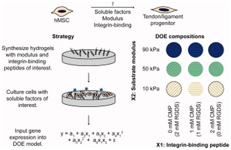
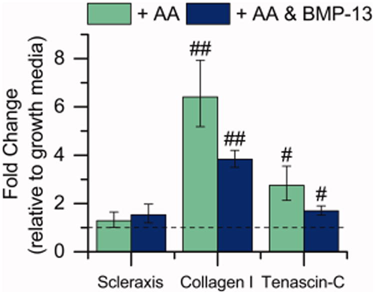
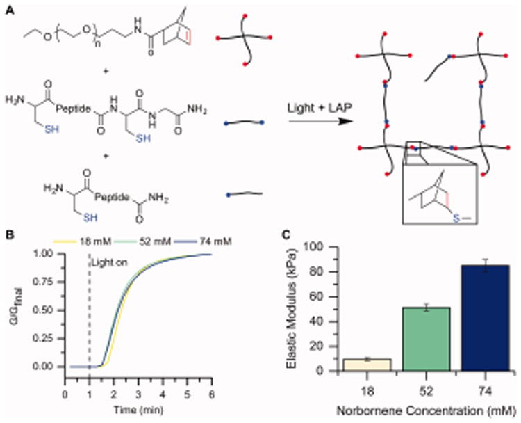
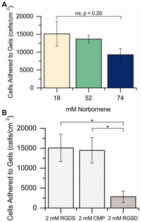
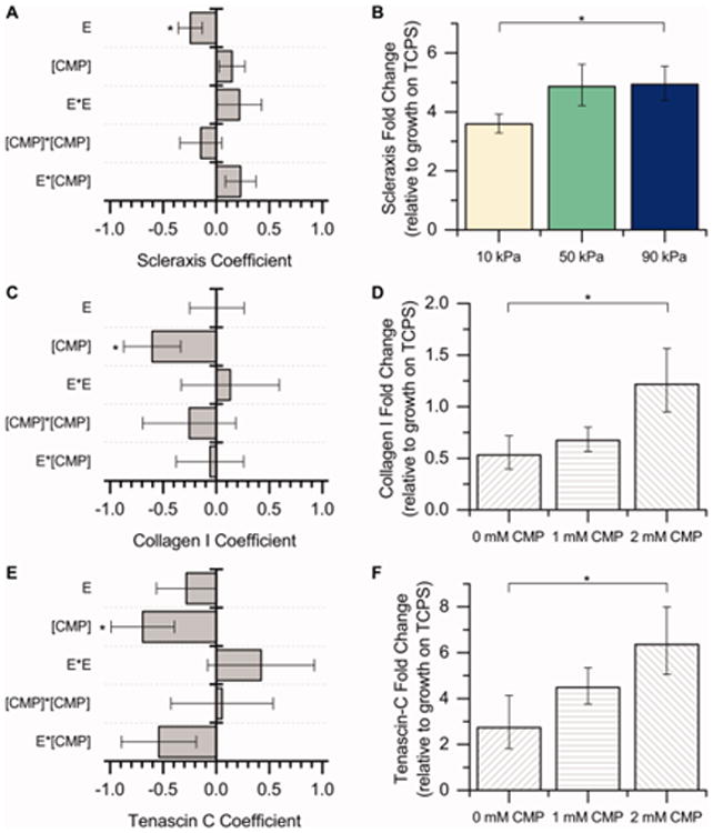
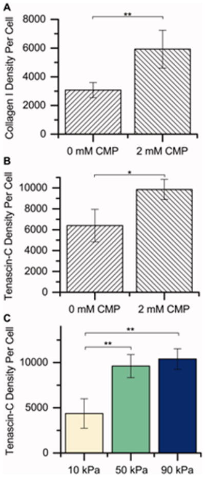
Mesenchymal stem cells (MSCs) are promising for the regeneration of tendon and ligament tissues. Toward realizing this potential, microenvironment conditions are needed for promoting robust lineage-specific differentiation into tenocytes/ligament fibroblasts. Here, we utilized a statistical design of experiments approach to examine combinations of matrix modulus, composition, and soluble factors in human MSC tenogenic/ligamentogenic differentiation. Specifically, well-defined poly(ethylene glycol)-based hydrogels were synthesized using thiol-ene chemistry providing a bioinert base for probing cell response to extracellular matrix cues. Monomer concentrations were varied to achieve a range of matrix moduli (E ∼ 10-90 kPa), and different ratios of integrin-binding peptides were incorporated (GFOGER and RGDS for collagen and fibronectin, respectively), mimicking aspects of developing tendon/ligament tissue. A face-centered central composite response surface design was utilized to understand the contributions of these cues to human MSC differentiation in the presence of soluble factors identified to promote tenogenesis/ligamentogenesis (BMP-13 and ascorbic acid). Increasing modulus and collagen mimetic peptide content increased relevant gene expression and protein production or retention (scleraxis, collagen I, tenascin-C). These findings could inform the design of materials for tendon/ligament regeneration. More broadly, the design of experiments enabled efficient data acquisition and analysis, requiring fewer replicates than if each factor had been varied one at a time. This approach can be combined with other stimuli (for example, mechanical stimulation) toward a better mechanistic understanding of differentiation down these challenging lineages.

Keywords: cell microenvironment; design of experiments; hydrogel; mesenchymal stem cell; tendon.


Figures

Figure 1
Figure 1
Approach. The purpose of this study was to test the hypothesis that a combination of soluble factors, modulus, and integrin-binding moieties could induce hMSC differentiation into tenocytes/ligament fibroblasts. To study the effects of modulus and integrin-binding peptides on differentiation, well-defined PEG-based hydrogels were synthesized to mimic aspects of developing tendon/ligament, seeded with cells, and cultured in the presence of tendon-inducing soluble factors. A design of experiments approach was applied to simultaneously determine the individual and synergistic effects of the two input factors, a CMP integrin-binding peptide (×1) and substrate modulus (×2). The ΔΔCt values from gene expression measurements were fit as responses in a DOE model, as tenogenic gene expression is generally considered one of the most conclusive markers of tenogenic differentiation. Coefficients a1, a2, a3, a4, a5, a6, and ε were determined in Minitab 17.
Figure 2
Figure 2
Effect of soluble factors on expression of tendon-associated genes. The gene expression of hMSCs cultured on TCPS cultured with AA (50 μg/mL) and BMP-13 (10 ng/mL) was evaluated, in part to determine a baseline level of expression in the presence of soluble factors for comparison to effects in the presence of matrix mimics. Gene expression was measured after 15 days of culture. Significant upregulation of the tendon-associated genes collagen I and tenascin-C were observed [n = 10 for the growth samples, n = 13 for AA and AA and BMP-13 samples, #p < 0.05, ##p < 0.01 compared with growth medium].
Figure 3
Figure 3
Hydrogel formation and characterization. A) Hydrogels were formed by reaction of PEG-4Nb (chemical structure of one arm shown here), a dithiol crosslinking peptide (CGKGWGKGCG), and monofunctional bioactive or scrambled tethers. Upon addition of a photoinitiator (LAP) and light (365 nm), these macromers react by a thiol–ene reaction to form a water-stable bond and a hydrogel. B) Gelation was measured for the compositions of interest by forming the gels in situ on a rheometer. The change in modulus, as a measure of crosslink density, was monitored over time upon light exposure (5 mW/cm2 at 365 nm) to assess the progress of gelation. Here, the modulus is normalized to the final modulus. Each condition was observed to reach 95% of its final modulus value by 3 min after polymerization commenced (that is, 4 min after the run started), and consequently, 3 min of irradiation was chosen as the gelation time for subsequent experiments. Representative curves are shown for each condition. C) The Young's modulus (E) was determined by rheometry and rubber elasticity theory. As the concentration of PEG-norbornene increases (noted here by the total norbornene concentration), the modulus of the gel also increases. Conditions were identified to achieve moduli of E = 10 kPa, 50 kPa, and 90 kPa, as desired for the design of experiments model [n = 6].
Figure 4
Figure 4
hMSC attachment to substrates. A) hMSCs were seeded on hydrogels with different PEG-norbornene concentrations containing 2 mM RGDS, allowed to adhere for 24 h in growth media (10% FBS), and then starved for 24 h (1% FBS). After starvation, cells were counted to establish the initial seeding density (that is, the number of hMSCs on each hydrogel surface at day 0 of the differentiation study). The initial cell seeding density was statistically equal for all concentrations of PEG-norbornene tested in this study [n = 3 for 18 mM and 52 mM; n = 4 for 74 mM]. B) Similarly, the effect of varying the identity of the peptide tether incorporated into the hydrogels was assessed on 18 mM Nb substrates. No differences in initial cell seeding density were observed between the two bioactive sequences (RGDS and CMP), whereas the scrambled RGSD permitted significantly less hMSC adhesion to the hydrogel at day 0, suggesting some specificity in cell interactions with these synthetic ECMs [n = 3, * p < 0.05 by two-sample t test].
Figure 5
Figure 5
DOE analysis of microenvironment cues on tenogenic differentiation. A response surface methodology was applied to study the effect of modulus (E) and integrin-binding peptides ([CMP]) on hMSC tenogenic gene expression (scleraxis, collagen I, tenascin-C) after culture for 15 days in the presence of soluble factors (BMP-13 and AA). The coefficients for the DOE model [a2–a6 in Eq. (3)] are plotted here (left). The response variable into the model was ΔΔCt, so a negative coefficient value corresponds to an increase in expression. [*p < 0.05 indicates significance of the marked term by ANOVA.] A) Scleraxis gene expression is significantly impacted by hydrogel modulus, while C) collagen I and E) tenascin-C gene expression are significantly impacted by the presence of the CMP integrin-binding peptide. Conditions subsequently were binned to further understand the effects of increasing modulus or CMP alone (right). B) Since the only significant term for the scleraxis coefficient model was modulus, the overall fold change data are presented here for all integrin-binding peptides as a function of modulus. Increasing the hydrogel modulus from 10 kPa to 90 kPa led to significant upregulation of scleraxis in hMSCs. D, F) Since the only significant term for the collagen I and tenascin-C coefficient models was CMP, the overall fold change data are presented here for all moduli as a function of CMP concentration. Increasing the CMP concentration from 0 mM to 2 mM led to significant upregulation of collagen I and tenascin-C in hMSCs. These results suggest that both increasing modulus and increasing CMP concentration increase hMSC tenogenic gene expression, an indicator of tenogenic differentiation [n = 7 for each condition, *p < 0.05 by Fisher's post hoc test].
Figure 6
Figure 6
Semiquantitative analysis of protein content by immunocytochemistry. To determine if differences in gene expression resulted in observable differences in protein production, we performed a semiquantitative analysis of protein expression for a subset of samples (each modulus with 0 or 2 mM CMP [2 or 0 mM RGDS, respectively]). Here, we immunostained for collagen I and tenascin-C, imaged the samples, and summed the fluorescence intensities (raw integrated density in ImageJ) in each image, normalizing to the number of nuclei. The factors (CMP and modulus) were analyzed individually to determine their effects on protein expression since the interaction terms (CMP × modulus) by ANOVA were not statistically significant (see Table I). Increasing the CMP concentration from 0 mM CMP (2 mM RGDS) to 2 mM CMP (0 mM RGDS) significantly increased the staining intensity for A) collagen I and B) tenascin-C, indicating increased protein expression, consistent with gene expression observations. C) Increasing the modulus of the substrate from 10 kPa to 50 kPa or 90 kPa also significantly increased the staining intensity for tenascin-C [n = 3 for each condition, *p < 0.05 by Fisher's post hoc test, **p < 0.01 by Fisher's post hoc test].
Figure 7
Figure 7
Lineage specificity in controlled microenvironment. hMSCs were cultured on 90 kPa and 2 mM CMP gels with BMP-13 and AA (the condition observed to most significantly affect tenogenic gene expression in the DOE), or in growth media on TCPS (control), for 15 days; expression of genes associated with nontenogenic lineages was measured with a PCR plate. Significant downregulation of the adipogenic gene CEBPA, the osteogenic genes ALP and RUNX2 (early markers), and the chondrogenic gene ACAN were observed. Significant upregulation of the osteogenic gene BGLAP was observed. Thus, the combinations of factors presented here did not lead to significant degrees of adipogenesis or chondrogenesis, but a high modulus and high CMP substrate, in combination with the soluble factors AA and BMP-13, led to an increase in the late osteogenic marker BGLAP [n = 3, **p < 0.01 compared with growth media on TCPS].

Similar articles

Cited by

References

    1. September AV, Schwellnus MP, Collins M. Tendon and ligament injuries: the genetic component. Br J Sports Med. 2007;41:241–6. - PMC - PubMed
    1. Chahal J, Lee A, Heard W, Bach BR. A Retrospective Review of Anterior Cruciate Ligament Reconstruction Using Patellar Tendon: 25 Years of Experience. Orthop J Sport Med. 2013;1:2325967113501789. - PMC - PubMed
    1. Kartus J, Movin T, Karlsson J. Donor-site morbidity and anterior knee problems after anterior cruciate ligament reconstruction using autografts. Arthroscopy. 2001;17:971–80. - PubMed
    1. Fleming BC, Hulstyn MJ, Oksendahl HL, Fadale PD. Ligament Injury, Reconstruction and Osteoarthritis. Curr Opin Orthop. 2005;16:354–62. - PMC - PubMed
    1. Rodrigues MT, Reis RL, Gomes ME. Engineering tendon and ligament tissues: present developments towards successful clinical products. J Tissue Eng Regen Med. 2013;7:673–86. - PubMed

Publication types