. 2016 Apr;147(4):453-63.
doi: 10.1111/imm.12578. Epub 2016 Feb 8.

The atypical IκB protein IκB(NS) is important for Toll-like receptor-induced interleukin-10 production in B cells

Affiliations

The atypical IκB protein IκB(NS) is important for Toll-like receptor-induced interleukin-10 production in B cells

Minami Miura et al. Immunology. 2016 Apr.

Abstract

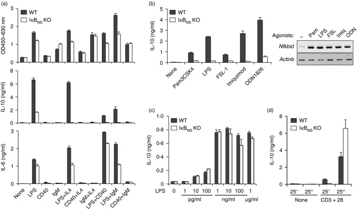
Although a major function of B cells is to mediate humoral immunity by producing antigen-specific antibodies, a specific subset of B cells is important for immune suppression, which is mainly mediated by the secretion of the anti-inflammatory cytokine interleukin-10 (IL-10). However, the mechanism by which IL-10 is induced in B cells has not been fully elucidated. Here, we report that IκBNS , an inducible nuclear IκB protein, is important for Toll-like receptor (TLR)-mediated IL-10 production in B cells. Studies using IκB(NS) knockout mice revealed that the number of IL-10-producing B cells is reduced in IκB(NS)(-/-) spleens and that the TLR-mediated induction of cytoplasmic IL-10-positive cells and IL-10 secretion in B cells are impaired in the absence of IκB(NS). The impairment of IL-10 production by a lack of IκB(NS) was not observed in TLR-triggered macrophages or T-cell-receptor-stimulated CD4(+) CD25(+) T cells. In addition, IκB(NS)-deficient B cells showed reduced expression of Prdm1 and Irf4 and failed to generate IL-10(+) CD138(+) plasmablasts. These results suggest that IκB(NS) is selectively required for IL-10 production in B cells responding to TLR signals, so defining an additional role for IκB(NS) in the control of the B-cell-mediated immune responses.

Keywords: IκBNS; Toll-like receptors; interleukin-10-producing B cells.


Figures

Figure 1
Figure 1
Iκ BNS −/− mice contain reduced numbers of interleukin‐10 (IL‐10)‐producing B cells in the spleen. (a) Flow cytometric analysis of CD1dhi CD5+ B cells in spleen from wild‐type (Iκ BNS +/+, WT) and Iκ BNS‐deficient (Iκ BNS −/−, KO) mice. Representative FACS profiles of CD19+ spleen B cells for CD1d and CD5 are shown. Numbers represent the percentage of cells in indicated gates. (b) The frequency of CD1dhi CD5+ cells in CD19+ splenic B cells (left) and the numbers of CD1dhi CD5+ B cells in wild‐type and Iκ BNS −/− spleens (right) are shown. (c) Flow cytometric analysis of IL‐10‐producing B cells in spleen. Splenocytes from wild‐type and Iκ BNS −/− mice were incubated with lipopolysaccharide (LPS) plus PMA, A23187 and GolgiPlug for 5 hr. The cells were then stained with anti‐CD19 and anti‐IL‐10. Representative dot plots show the frequencies of cytoplasmic IL‐10+ cells in CD19+ B cells. (d) The frequency of cytoplasmic IL‐10+ cells in CD19+ splenic B cells (left) and the number of IL‐10 producing B cells in the spleen (right) are shown. For (a) and (b), data are representative of at least three independent experiments. For (b) and (d), each symbol indicates an individual mouse. Horizontal bars represent the mean. n > 5; **P < 0·01.
Figure 2
Figure 2
B‐cell‐intrinsic deficiency in Iκ BNS results in the impaired generation of interleukin‐10 (IL‐10) ‐producing B cells in vivo. bone marrow (BM) cells from B6.μMT mice were mixed with BM cells from wild‐type or Iκ BNS −/− mice at a ratio of 4:1, and 1 × 10⁷ BM cells were transferred intravenously into irradiated B6.μMT mice. Lipopolysaccharide (LPS) ‐induced IL‐10 production in donor‐derived B cells was examined 8 weeks post transfer by the daily administration of LPS (10 μg, intraperitoneally) for 3 days. Controls received PBS alone. Two days after the last inoculum, splenocytes were stained with surface CD19 and intracellular IL‐10. (a) Representative FACS profiles of CD19+ splenocytes for the expression of cytoplasmic IL‐10 with the percentages of cells in gates. (b) The frequency of IL‐10‐producing cells in CD19+ splenic B cells. Data from four control and eight LPS‐treated mice are shown. Each symbol shows an individual mouse, and horizontal bars represent the mean. Two independent experiments were performed, and similar results were obtained. **P < 0·01.
Figure 3
Figure 3
Iκ BNS‐deficient B cells fail to produce lipopolysaccharide (LPS) ‐induced interleukin‐10 (IL‐10). (a) Splenic B cells from wild‐type and Iκ BNS −/− mice were cultured with various concentrations of LPS for 3 days. The cells were then FACS stained with anti‐CD19 and anti‐IL‐10. Representative FACS profiles of CD19+ B cells for cytoplasmic IL‐10 with the frequencies of cells in indicated gates are shown. (b) The amount of IL‐10 in the culture supernatant of LPS‐stimulated B cells. Splenic B cells were stimulated with LPS alone or LPS plus PMA, A23187 for 3 days. (c) Splenic B cells from wild‐type and Iκ BNS −/− mice were activated with LPS (5 μg/ml) for 1 day, and they were co‐cultured with wild‐type T cells under stimulation with anti‐CD3ε and anti‐CD28 for 2 days. The expression level of IL‐10 in B cells and the expression levels of IL‐10 and interferon‐γ (IFNγ) in CD3ε + T cells were analysed by flow cytometry. Representative FACS plots with the frequencies of cells in indicated gates are shown. The bar graph shows the frequencies of IFNγ + cells in CD3ε + T cells (dot columns), IL‐10+ cells in CD19+ B cells (filled columns), and IL‐10+ cells in CD3ε + T cells (open columns) in the co‐culture. *P < 0·05, **P < 0·01. (d) Purified CD1dhi CD5+ B cells from wild‐type and Iκ BNS −/− spleens were incubated with or without LPS for 2 days, and the amount of IL‐10 in the culture supernatant was measured by ELISA. (e) Purified CD21hi CD23lo marginal zone (MZ) B cells and CD21+ CD23hi follicular B cells from wild‐type and Iκ BNS −/− spleens were incubated with LPS (5 μg/ml) for 4 days, and the amount of IL‐10 in the culture supernatant was measured by ELISA. The data shown were obtained from triplicate assay and represent at least two independent experiments. Error bars represent the standard deviation.
Figure 4
Figure 4
Toll‐like receptor (TLR) ‐induced interleukin‐10 (IL‐10) production is impaired in Iκ BNS‐deficient B cells. (a) Splenic B cells isolated from wild‐type and Iκ BNS −/− spleens were incubated for 2 days in the presence of the indicated reagents [5 μg/ml lipopolysaccharide (LPS), 5 μg/ml anti‐CD40, 10 μg/ml anti‐IgM, 10 ng/ml rIL‐4]. The proliferative activity of B cells was measured using a cell counting kit‐8 (top). Culture supernatant was used for ELISA to determine the amounts of IL‐10 (middle) and IL‐6 (bottom). (b) Purified splenic B cells were cultured with various TLR agonists for 2 days to collect supernatants or for 2 hr for the RTPCR analysis. The semi‐quantitative RTPCR analysis shows the TLR‐induced expression of Nfkbid in wild‐type B cells. β‐Actin was used as a loading control. The concentration of each TLR agonist is described in the Materials and methods. (c) Naive macrophages in the peritoneal cavity were collected from wild‐type and Iκ BNS −/− mice and stimulated with the indicated amounts of LPS for 2 days. (d) CD4+ CD25 conventional T cells (25) and CD4+ CD25+ regulatory T cells (25+) from wild‐type and Iκ BNS −/− lymph nodes were FACS sorted and stimulated with or without anti‐CD3ε and anti‐CD28 for 3 days. The amount of cytokines in the culture supernatants was determined by a triplicate ELISA. Data represent at least two independent experiments.
Figure 5
Figure 5
Expression of cytokine genes and nuclear factors in lipopolysaccharide (LPS) ‐activated Iκ BNS‐deficient B cells. (a, b) The LPS‐induced transcription of Il10 is reduced in Iκ BNS‐deficient B cells during the first 24 hr of induction. Total RNA was isolated from purified splenic B cells stimulated with 5 μg/ml of LPS for the indicated periods, and the semi‐quantitative RTPCR assessment of cytokine gene transcripts was performed. The LPS‐induced transcription kinetics of Nfkbid, Il10 and Il6 genes (a) and cytokine expression profiles within 24 hr (b) are shown. (c) The expression and nuclear localization of Iκ BNS, NFκBp65, and p50 were analysed based on the immunoblotting of nuclear extracts prepared from LPS‐stimulated wild‐type and Iκ BNS −/− B cells. The nuclear extracts were also used for DNA pull down with biotinylated probes for two interleukin‐10 (IL‐10) promoters containing a putative nuclear factor‐κB (NFκB)‐binding site. (d) Western blot analysis of DNA pull down for the nuclear lysates and IL‐10‐promoter probes containing proximal (−55/−46) or distal (−861/−851) NFκB motif. (e) The expression of NFATc1 was analysed based on the immunoblotting of nuclear extracts prepared from LPS‐stimulated wild‐type and Iκ BNS −/− B cells. Iκ BNS shows two and NFATc1 shows several bands depending on their phosphorylation status. Lamin B was used as a nuclear loading control. All data represent at least two independent experiments.
Figure 6
Figure 6
Toll‐like receptor (TLR) ‐induced differentiation of IL‐10+ CD138+ plasmablasts is impaired in the absence of Iκ BNS. (a, b) Real‐time PCR analysis of the expression of Prdm1 and Irf4. FACS‐sorted CD1dhi CD5+ B cells (a) and CD21hi CD23lo MZ B cells (b) from wild‐type and Iκ BNS −/− spleens were cultured for the indicated periods in the presence of 5 μg/ml lipopolysaccharide (LPS), and total RNA was isolated and subjected to quantitative PCR analysis. All expression data were normalized to β‐actin expression. The data are representative of two independent experiments. *P < 0·05, **P < 0·01. (c) Splenic B cells isolated from wild‐type and Iκ BNS −/− mice were stimulated with LPS (1·0 μg/ml) for 2 or 3 days, and the expression levels of surface CD138, CD44, and cytoplasmic interleukin 10 (IL‐10) on CD19+ B cells were analysed by flow cytometry. (d) Splenic B cells from wild‐type and Iκ BNS −/− mice were incubated with various TLR agonists for 3 days, and the expression of CD138 and cytoplasmic IL‐10 were analysed by flow cytometry. Representative FACS profiles with the percentages of cells in quadrant gates are shown. The data represent three independent experiments.

References

    1. Bekeredjian‐Ding I, Jego G. Toll‐like receptors − sentries in the B‐cell response. Immunology 2009; 128:311–23. - PMC - PubMed
    1. Booth J, Wilson H, Jimbo S, Mutwiri G. Modulation of B cell responses by Toll‐like receptors. Cell Tissue Res 2011; 343:131–40. - PubMed
    1. Bernasconi NL, Onai N, Lanzavecchia A. A role for Toll‐like receptors in acquired immunity: up‐regulation of TLR9 by BCR triggering in naive B cells and constitutive expression in memory B cells. Blood 2003; 101:4500–4. - PubMed
    1. Ruprecht CR, Lanzavecchia A. Toll‐like receptor stimulation as a third signal required for activation of human naive B cells. Eur J Immunol 2006; 36:810–16. - PubMed
    1. Peng SL. Signaling in B cells via Toll‐like receptors. Curr Opin Immunol 2005; 17:230–6. - PubMed

Publication types

MeSH terms

LinkOut - more resources