. 2016 Jan 7;98(1):75-89.
doi: 10.1016/j.ajhg.2015.11.018. Epub 2015 Dec 31.

Autosomal-Dominant Corneal Endothelial Dystrophies CHED1 and PPCD1 Are Allelic Disorders Caused by Non-coding Mutations in the Promoter of OVOL2

Affiliations

Autosomal-Dominant Corneal Endothelial Dystrophies CHED1 and PPCD1 Are Allelic Disorders Caused by Non-coding Mutations in the Promoter of OVOL2

Alice E Davidson et al. Am J Hum Genet. .

Abstract

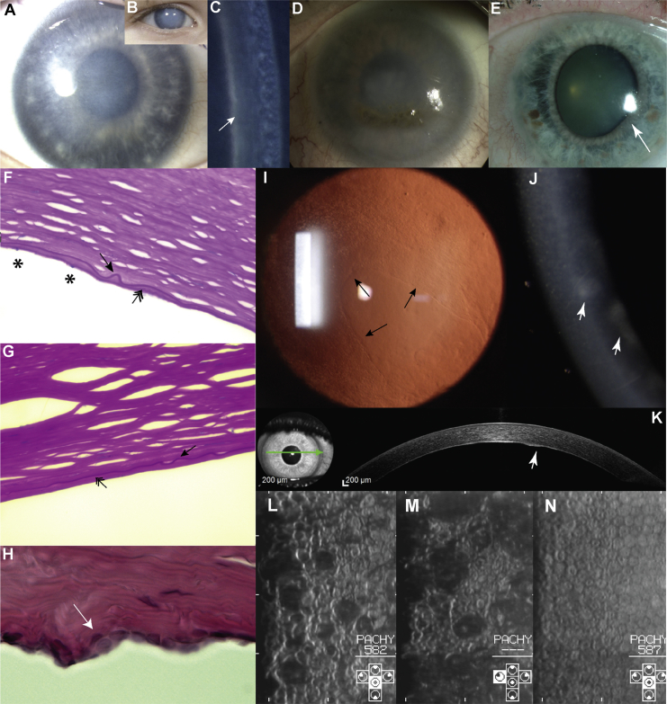
Congenital hereditary endothelial dystrophy 1 (CHED1) and posterior polymorphous corneal dystrophy 1 (PPCD1) are autosomal-dominant corneal endothelial dystrophies that have been genetically mapped to overlapping loci on the short arm of chromosome 20. We combined genetic and genomic approaches to identify the cause of disease in extensive pedigrees comprising over 100 affected individuals. After exclusion of pathogenic coding, splice-site, and copy-number variations, a parallel approach using targeted and whole-genome sequencing facilitated the identification of pathogenic variants in a conserved region of the OVOL2 proximal promoter sequence in the index families (c.-339_361dup for CHED1 and c.-370T>C for PPCD1). Direct sequencing of the OVOL2 promoter in other unrelated affected individuals identified two additional mutations within the conserved proximal promoter sequence (c.-274T>G and c.-307T>C). OVOL2 encodes ovo-like zinc finger 2, a C2H2 zinc-finger transcription factor that regulates mesenchymal-to-epithelial transition and acts as a direct transcriptional repressor of the established PPCD-associated gene ZEB1. Interestingly, we did not detect OVOL2 expression in the normal corneal endothelium. Our in vitro data demonstrate that all four mutated OVOL2 promoters exhibited more transcriptional activity than the corresponding wild-type promoter, and we postulate that the mutations identified create cryptic cis-acting regulatory sequence binding sites that drive aberrant OVOL2 expression during endothelial cell development. Our data establish CHED1 and PPCD1 as allelic conditions and show that CHED1 represents the extreme of what can be considered a disease spectrum. They also implicate transcriptional dysregulation of OVOL2 as a common cause of dominantly inherited corneal endothelial dystrophies.


Figures

Figure 1
Figure 1
Pedigree Structure of CHED1- and PPCD1-Affected Families with OVOL2 Promoter Mutations Pedigrees of (A) the index British CHED1-affected family (BR1), (B) 16 PPCD1-affected families (C1–C14, C25, and C30) from the Czech Republic, and further British families (C) BR2 and (D) BR3. For each family (BR1–BR3) or group of families (C1–C14, C25, and C30), a distinct and unique mutation in the OVOL2 promoter was identified. Sanger sequencing traces representing the four unique heterozygous mutations identified are shown in each respective panel (A–D). Second-degree unaffected relatives are not shown. Mutations are annotated in accordance with GenBank: NM_021220 (Ensembl: ENST00000278780), and +1 represents the A of the ATG translation initiation codon. DNA from individuals, each highlighted with a red arrow, was analyzed by targeted re-sequencing of the disease-associated locus (chr20: 17,335,789–19,665,902). Asterisks indicate that a DNA sample was available for genotyping. Abbreviations are as follows: W, whole-exome sequencing (WES); and pw, pooled WES. Dashed lines indicate that a sample was whole-genome sequenced.
Figure 2
Figure 2
Spectrum of Corneal Disease Associated with OVOL2 Mutations (A–C) Individual VII:13 from family BR1 at age 3 years. The right (A) and left (B) corneas have a hazy appearance on direct illumination and an increased thickness from diffuse corneal edema. (C) A prominent Descemet membrane can be seen with narrow-beam illumination (arrow). (D) Individual VI:2 from family BR1 at age 52 years (without surgery) shows secondary lipoidal degeneration. (E) Individual III:3 from family BR3 shows a clear cornea but a distorted iris (arrow) secondary to the presence of a peripheral area of iris-to-corneal adhesion (not shown). (F–H) Histological specimens. (F) Individuals VII:13 (age 6 years) and (G) VII:7 (age 11 years) from family BR1. Both individuals have a thin and irregular Descemet membrane (arrow), reduced endothelial cell count (asterisks), and accumulation of material posterior to the Descemet membrane, possibly reflecting mild retrocorneal fibrosis (double-headed arrow). (H) Individual III:1 from family C11 (42 years) shows focal multilayering of endothelial cells (arrow) and undulation of the posterior corneal surface. (I) Retroillumination image of the cornea of individual II:1 (age 29 years) from family C30 shows a mild presentation. An island of normally appearing endothelial cells is surrounded by abnormal cells (arrows highlight the boundary of cells with a normal appearance). (J and K) Individual III:5 (age 33 years) from family C3. (J) A narrow-beam section of the right cornea demonstrates a slight corneal haze and gray focal areas at the level of the Descemet membrane and endothelium (arrowhead). (K) An ocular-coherence-tomography cross-section of the cornea demonstrates a raised lesion on the posterior corneal surface (arrowhead). (L and M) Corneal endothelial specular images from individual V:11 (age 13 years) from family C2. Note the variation in the individual size and shape of the endothelial cells. The dark areas presumably correspond to elevated regions. (N) For comparison, the corneal endothelial specular image of a 25-year-old unaffected sister of individual V:11 (V:10 from family C2) shows a normal appearance of cells with a uniform size and shape.
Figure 3
Figure 3
Schematic Representation of the Overlapping Loci, Conservation of the OVOL2 Promoter, and Relative Position of the Four Mutations (A) Overlapping disease-associated loci on 20p for the Czech familial cohort and the refined region in family BR1 (Figure S1). (B) Annotated transcripts within the linked regions. (C) Schematic illustration of OVOL2, which comprises four exons and encodes a 275 aa protein. (D) ClustalW2 multiple alignment of the promoter region of 11 OVOL2 orthologs indicates the position of the four mutations identified. The following orthologs were used for the alignment: Homo sapiens (Ensembl: ENSG00000125850), Pan troglodytes (Ensembl: ENSPTRG00000013280), Pongo abelii (Ensembl: ENSPPYG00000010732), Callithrix jacchus (Ensembl: ENSCJAG00000005049), Mus musculus (Ensembl: ENSMUSG00000037279), Rattus norvegicus (Ensembl: ENSRNOG00000006850), Canis familiaris (Ensembl: ENSCAFG00000005453), Equus caballus (Ensembl: ENSECAG00000007899), Pteropus vampyrus (Ensembl: ENSPVAG00000001140), Dasypus novemcinctus (Ensembl: ENSDNOG00000025067), Monodelphis domestica (Ensembl: ENSMODG00000005504), and Xenopus tropicalis (Ensembl: ENSXETG00000024897). The alignment illustrates the conservation of a 145 bp promoter region in which the four disease-associated mutations were identified (mutated base pairs are highlighted in red and boxed). The transcription start site is indicated with an arrow. Mutations are annotated in accordance with the OVOL2 cDNA sequence (GenBank: NM_021220; Ensembl: ENST00000278780), and +1 represents the A of the ATG translation initiation codon.
Figure 4
Figure 4
Functional Analysis of the OVOL2 Promoter (A) OVOL2 mutations are located within a transcriptionally active region of the promoter. Interrogation of publically available ChIP-seq data released as part of the ENCODE project demonstrates that multiple transcription factors, including FOXA1, FOXA2, NRF1, SP1, CTBP2, and EP300, bind the region encompassing all OVOL2 disease-associated mutations. (B) Mutations in the OVOL2 promoter cause an increase in gene expression in vitro. A dual-luciferase reporter assay was used for assessing the impact of disease-associated mutations on the activity of the OVOL2 promoter. HEK293 cells were co-transfected with pRL-CMV (Renilla luciferase) and pGL3-Basic (firefly luciferase) containing 1,824 bp of the wild-type or respective mutant OVOL2 promoter sequence. The ratio of firefly to Renilla luciferase activity was calculated for all samples. Wild-type data were normalized to 1, and the relative luciferase activity in all other samples is expressed in relation to the wild-type data. All mutations investigated significantly increased the relative luciferase activity. Data represent a minimum of three independent experiments with triplicate measurements. Error bars represent ± 1 SD. p values were calculated by one-way ANOVA (∗∗∗p ≤ 0.001).

References

    1. Weiss J.S., Møller H.U., Aldave A.J., Seitz B., Bredrup C., Kivelä T., Munier F.L., Rapuano C.J., Nischal K.K., Kim E.K. IC3D classification of corneal dystrophies--edition 2. Cornea. 2015;34:117–159. - PubMed
    1. Cibis G.W., Krachmer J.A., Phelps C.D., Weingeist T.A. The clinical spectrum of posterior polymorphous dystrophy. Arch. Ophthalmol. 1977;95:1529–1537. - PubMed
    1. Krachmer J.H. Posterior polymorphous corneal dystrophy: a disease characterized by epithelial-like endothelial cells which influence management and prognosis. Trans. Am. Ophthalmol. Soc. 1985;83:413–475. - PMC - PubMed
    1. Pearce W.G., Tripathi R.C., Morgan G. Congenital endothelial corneal dystrophy. Clinical, pathological, and genetic study. Br. J. Ophthalmol. 1969;53:577–591. - PMC - PubMed
    1. Yellore V.S., Papp J.C., Sobel E., Khan M.A., Rayner S.A., Farber D.B., Aldave A.J. Replication and refinement of linkage of posterior polymorphous corneal dystrophy to the posterior polymorphous corneal dystrophy 1 locus on chromosome 20. Genet. Med. 2007;9:228–234. - PubMed

Publication types

Supplementary concepts