. 2016 Apr;37(4):1349-60.
doi: 10.1002/hbm.23106. Epub 2016 Jan 9.

FMRI of spinal and supra-spinal correlates of temporal pain summation in fibromyalgia patients

Affiliations

FMRI of spinal and supra-spinal correlates of temporal pain summation in fibromyalgia patients

Rachael L Bosma et al. Hum Brain Mapp. 2016 Apr.

Abstract

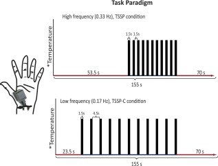
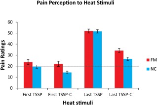
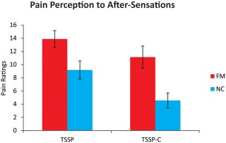
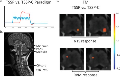
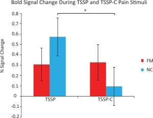
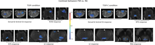
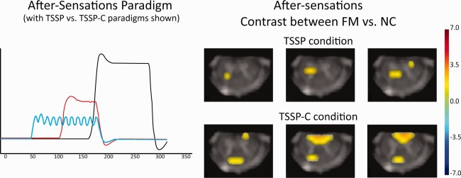
Fibromyalgia syndrome (FM) is a debilitating chronic pain condition, which afflicts primarily females. Although the etiology of this illness is not completely understood, FM pain is thought to rely on enhanced pain sensitivity maintained by central mechanisms. One of these mechanisms is central pain amplification, which is characterized by altered temporal summation of second pain (TSSP). Here we use a TSSP paradigm and functional MRI (fMRI) of the spinal cord, brainstem, and brain to noninvasively examine the central nervous system contributions to TSSP in FM patients and normal controls (NC). Functional MRI of pain-free female adults (N = 15) and FM patients (N = 14) was conducted while brief, repetitive heat pain stimuli (0.33 Hz) were applied to the thenar eminence of the hand (C6 dermatome). The stimulus intensity was adjusted to each participant's heat pain sensitivity to achieve moderate pain. Data were analyzed by means of a General Linear Model and region-of-interest analyses. All participants demonstrated significant pain summation in the TSSP condition. FM subjects, however, required significantly lower stimulus intensities than NC to achieve similar TSSP. fMRI analyses of perceptually equal TSSP identified similar brain activity in NC and FM subjects; however, multiple areas in the brainstem (rostral ventromedial medulla and periaqueductal grey region) and spinal cord (dorsal horn) exhibited greater activity in NC subjects. Finally, increased after-sensations and enhanced dorsal horn activity was demonstrated in FM patients. In conclusion, the spinal and brainstem BOLD responses to TSSP are different between NC and FM patients, which may indicate alterations to descending pain control mechanisms suggesting contributions of these mechanisms to central sensitization and pain of FM patients.

Keywords: brainstem; fMRI; fibromyalgia; spinal cord; temporal summation of pain.


Figures

Figure 1
Figure 1
The task paradigm for the TSSP and TSSP‐C conditions. The temperature of stimulation was calibrated for each individual to produce a moderate pain rating (50 on a 100‐point scale) for the last stimulus of the TSSP paradigm. The same temperature was used for both conditions. The duration of the first baseline period was reduced for the TSSP‐C condition, so that the final baseline period was the same between conditions. For the TSSP condition, the heat stimuli were applied every 3 s, for 1.5 s, whereas in the TSSP‐C condition, the heat stimuli were applied every 6 s, also for a duration of 1.5 s. [Color figure can be viewed in the online issue, which is available at http://wileyonlinelibrary.com.]
Figure 2
Figure 2
The mean pain ratings across participants for the first and last heat pain stimulus in the two conditions. In the fMRI session, all participants were asked to remember the number ratings of the pain intensity related to the first and last heat stimuli (11 heat stimuli in total) and reported their ratings at the end of the stimulation period. In both the NC and FM participants, the rating to the last heat stimulus was significantly greater than the rating to the first (p < 0.001). The ratings to the last heat stimulus was also significantly different between conditions (TSSP > TSSP‐C) (p < 0.001). A rating of 20 represents the pain threshold (dotted line). [Color figure can be viewed in the online issue, which is available at http://wileyonlinelibrary.com.]
Figure 3
Figure 3
Reported ratings of after‐sensations obtained from each participant, for each condition, during the fMRI training sessions. Ratings of residual sensations/pain on the hand were obtained 15 and 30 s after the last heat stimulus and were averaged for each participant. The ratings of the FM participants were significantly higher (FM > NC) (p < 0.05). In the FM group, there was no significant difference between the ratings in the TSSP condition compared to the TSSP‐C condition. [Color figure can be viewed in the online issue, which is available at http://wileyonlinelibrary.com.]
Figure 4
Figure 4
Results of the contrast analysis comparing BOLD signal changes between the TSSP and TSSP‐C conditions in the FM group. (a) On the left is an illustration of the task paradigms convolved with the hemodynamic response function as used in the GLM analysis (red = TSSP paradigm, blue = TSSP‐C). (b) A midline sagittal slice from the functional data of one participant is shown for reference and illustrates the approximate location of the midbrain, pons, medulla, and C6 cord segment. (c) Key areas with significantly different responses to the stimulation paradigm are demonstrated in the vicinity of the NTS and RVM in the medulla. The results are overlaid on high resolution transverse slices. The color scale indicates the significance of areas with different responses between the conditions. [Color figure can be viewed in the online issue, which is available at http://wileyonlinelibrary.com.]
Figure 5
Figure 5
The % signal change from the C6 dorsal horn during task stimulation in each condition (TSSP vs TSSP‐C) and each group (FM and NC). Error bars indicate standard error, p < 0.05. [Color figure can be viewed in the online issue, which is available at http://wileyonlinelibrary.com.]
Figure 6
Figure 6
Results of the contrast analysis comparing BOLD signal changes between the FM and NC groups in both the TSSP and TSSP‐C conditions. On the left is the contrast between groups for the TSSP condition. Key areas with significantly different responses to the stimulation paradigm are demonstrated in the vicinity of the dorsal and ventral horns, RVM and NTS in the medulla, and LC in the pons. Contrast of the TSSP‐C condition between groups (right) reveals differences in the dorsal and ventral horns, NTS in the medulla, LC in the pons, and PAG in the midbrain. [Color figure can be viewed in the online issue, which is available at http://wileyonlinelibrary.com.]
Figure 7
Figure 7
Key areas with significantly different responses to the after‐sensations between FM and NC are shown to be the right dorsal horn region of the C6 segment in both the TSSP and TSSP‐C conditions. On the left is an illustration of the after‐sensation paradigms convolved with the hemodynamic response function as used in the GLM analysis (red = TSSP paradigm, blue = TSSP‐C). [Color figure can be viewed in the online issue, which is available at http://wileyonlinelibrary.com.]

Similar articles

Cited by

References

    1. Baliki MN, Chialvo DR, Geha PY, Levy RM, Harden RN, Parrish TB, Apkarian AV (2006): Chronic pain and the emotional brain: specific brain activity associated with spontaneous fluctuations of intensity of chronic back pain. J Neurosci 26:12165–12173. - PMC - PubMed
    1. Beck AT, Steer RA, Ball R, Ranieri W (1996): Comparison of Beck Depression Inventories ‐IA and ‐II in psychiatric outpatients. J Person Assess 67:588–597. - PubMed
    1. Bosma, RL , Ameli Mojarad, E , Leung, L , Pukall, C , Staud, R , Stroman, PW (2015): Neural correlates of temporal summation of second pain in the human brainstem and spinal cord. Hum Brain Mapp 36:5038–5050. - PMC - PubMed
    1. Bosma RL, Stroman PW (2014a): Assessment of data acquisition parameters, and analysis techniques for noise reduction in spinal cord fMRI data. Magn Reson Imaging 32:473–481. - PubMed
    1. Bosma, RL , Stroman, PW (2014b): Spinal cord response to stepwise and block presentation of thermal stimuli: A functional MRI study. J Magn Reson Imag JMRI 1318–1325. - PubMed

Publication types