. 2016 Jan 19;44(1):155-166.
doi: 10.1016/j.immuni.2015.11.004. Epub 2015 Dec 29.

Interleukin-2-Dependent Allergen-Specific Tissue-Resident Memory Cells Drive Asthma

Affiliations

Interleukin-2-Dependent Allergen-Specific Tissue-Resident Memory Cells Drive Asthma

Brian D Hondowicz et al. Immunity. .

Abstract

Exposure to inhaled allergens generates T helper 2 (Th2) CD4(+) T cells that contribute to episodes of inflammation associated with asthma. Little is known about allergen-specific Th2 memory cells and their contribution to airway inflammation. We generated reagents to understand how endogenous CD4(+) T cells specific for a house dust mite (HDM) allergen form and function. After allergen exposure, HDM-specific memory cells persisted as central memory cells in the lymphoid organs and tissue-resident memory cells in the lung. Experimental blockade of lymphocyte migration demonstrated that lung-resident cells were sufficient to induce airway hyper-responsiveness, which depended upon CD4(+) T cells. Investigation into the differentiation of pathogenic Trm cells revealed that interleukin-2 (IL-2) signaling was required for residency and directed a program of tissue homing migrational cues. These studies thus identify IL-2-dependent resident Th2 memory cells as drivers of lung allergic responses.


Figures

Figure 1
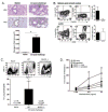
Figure 1. Detection of Derp1+ CD4+ T cells
A) Gating scheme to identify naïve or antigen-experienced Derp1+ CD4+ T cells in the SLO (top row) and lung (Thy1.2) (bottom row). B) Representative immunoflourescent sections of lungs from naïve (left panel) or day 3 post-challenge (right panel) mice showing the presence of CD4+ T cells only in the lungs of challenged mice (AW = airway and BV = blood vessel). These data were from one of two independent experiments from a total of 3 naïve and 3 challenged mice. See also Fig. S1.
Figure 2
Figure 2. Derp1+ specific cells produce Th2 type cytokines and cause inflammation
A) H&E and PAS stains of naïve or day 3 post challenge lungs. Top row shows the cellular infiltrate in sensitized lungs (right) compared to lungs from naïve mice (left). The PAS sections show glycogen (purple, right panel) production in lungs described above. Bar graph depicts the average (+/− S.D.) amount of glycogen in lungs of naïve or day 3 post challenge mice. The data shown are from 10 sections from two independent experiments with a total of 3 mice in each group and (*) (p<0.05) indicates statistical significance. B) Representative huCD2 (IL-4, left column) and IL-13 (middle column) plots from KN2 (IL-4) and IL-13eGFP reporter mice, respectively. Data are gated on CD4+ Derp1+ events acquired 3 days after challenge and were measured in 2–3 separate experiments. Bar graphs show the average percentage (+/− S.D.) of IL-4 and IL-13 in the SLO (top row) or lung parenchyma (bottom row). Data are the combined average from two independent experiments (n=5 for IL-4 expression, n=4 for IL-13 expression). C) Contour plots showing SSC+ Siglec F+ eosinophils from naïve WT or immunized WT or MHCII−/− mice (3 days post challenge). Graph shows the percentage of gated live cells from lungs that were eosinophils (CD3 Class II CD11b+ Siglec F+ SSC+). Data were collected from 2–9 separate experiments (naïve mice: n=4, WT challenged mice n=17, Class II KO challenged mice n=4). (*) indicates significantly different (p <0.05) compared to naïve and Class II KO mice. D) Graph shows the increase in airway resistance versus PBS control from naïve or immunized B6 and Class II KO mice. The data shown were collected from two experiments and the average +/− S.D. is graphed (naïve B6 n=4, immunized B6 n=4, naïve MHC class II KO n=3, immunized MHC Class II KO n=4) and (*) (p<0.05) indicates statistically different compared to all other groups. See also Fig. S2.
Figure 3
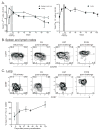
Figure 3. Derp1+ memory cells are primarily CCR7+ in the spleen and lymph nodes but CD69+ CCR7 in the lung
A) A timecourse of the number of CD4+ Derp1+ cells in the SLO (left, white circles), Thy1.2 Derp1+ cells in the lung (left, black circles), and total Thy1.2 CD4+ in lungs (right). Data for the graph were compiled from two experiments with 2–5 mice per timepoint. B) Representative contour plots of CCR7 and CXCR5 expression from CD4+ Derp1+ cells from the SLO. C) Representative contour plots of CCR7 and CD69 expression from Thy1.2 CD4+ Derp1+ cells from lungs. The graph shows the average percentage +/− S.D. of CD4+ Thy1.2 Derp1+ cells that are CD69+ at indicated timepoints in lungs. The grey column shows the time of allergic challenge. The represented data were collected from two experiments with 2–5 mice per timepoint. (*) indicates significantly higher percentage of CD69 compared to all other timepoints (p<0.05).
Figure 4
Figure 4. HDM immunization induces CD4+ CD69+ Derp1+ Trm in the lung
A) Cartoon summary of the parabiosis protocol. Dot plots demonstrate that i.v. anti-Thy1.2 antibody labeled all blood T cells and that both mice in the parabiont pair had roughly equal amounts of cells from naïve and immunized parabionts. B) Representative contour plots showing CD4+ Derp1+ cells from the SLO and lungs from immunized and naïve parabionts. The representative dot plots in the third column show that the resident CD4+ CD44+ Derp1+ are CD69+ and only found in the immunized parabiont. C) The graph indicates the number of CD69+ Derp1+ cells per mouse (n=8) found in lungs of naïve and immunized parabionts. The experiment was performed twice with 4 mice in each group and the (*) indicates that the two groups are significantly different (p<0.05).
Figure 5
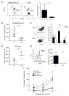
Figure 5. Resident lung cells are sufficient to induce AHR
A) Graph demonstrates the number of CD45.2+ (black circle) and CD4+ (white squares) in the blood before and during FTY720 administration. Each time point shows the average number +/− S.D. from 3 mice of the indicated cell population per μl/blood from one of two experiments. (*) significantly less CD45.2+ and CD4+ cells during FTY720 treatment compared to pre-treatment (p<0.05) B) Graph shows the increase in airway resistance to increasing doses of methacholine versus PBS. Each point on the graph is the average +/− S.D. airway resistance from 5 mice from two independent experiments. (*) indicates significantly greater airway resistance versus naïve B6 mice (p<0.05). C) The bar graphs indicate average +/− S.D number of CD4+ Derp1+ cells from 5 mice in SLO or lungs from control (black bars) or FTY720 treated (white bars) mice. Data were compiled from two experiments. D) Representative dot blot from two experiments showing CCR7 and CD69 expression on Thy1.2 CD4+ Derp1+ cells from lungs of control or FTY720 treated mice. See also Fig. S3.
Figure 6
Figure 6. Trm accumulation in the lungs is dependent on CD25
A) Representative dot plots from WT:CD25KO mixed bone marrow chimeric mice 6 days after various timepoints showing the percent of CD4+ CD44+ Derp1+ originating from WT versus CD25KO mice. Bar graphs depict the ratio of the percentage WT/CD25KO CD4+ CD44+ Derp1+ cells from 6 days post primary sensitization (n=4 from 3 separate experiments), day 3 post challenge (n=5 from 3 independent experiments), and 27 days post challenge (n=2 from 1 experiment). (*) indicates a significant difference between the indicated groups (p<0.05). B) Representative contour plots from WT:CD25KO mixed bone chimeras six days after primary sensitization showing CD62L or CD69 levels by CXCR5 expression on CD4+ Derp1+ in the SLO. Plots show percentage of Derp1+ cells that are CXCR5 CD62L (top) or the total percentage of CD69+ cells (bottom) from SLO of individual WT:CD25KO chimeric mice. Data are compiled from 7–8 mice from four independent experiments. (*) indicates a significant difference between the WT and CD25KO cells (p<0.05). C) CCR4 and CXCR3 expression were compared in CD4+ Derp1+ cells from WT and CD25KO cells in WT:CD25KO mixed BM chimeras. Contour plots depict the indicated chemokine receptor expression 6 days after primary sensitization from CD4+ Derp1+ WT or CD25KO cells. Both CCR4 and CXCR3 expression (*) (p<0.05) are significantly greater in WT Derp1+ cells versus CD25KO Derp1+ T cells. Data were compiled from 4 independent experiments with a total of 7 mice. See also Fig. S4–S6.
Figure 7
Figure 7. Trm accumulation in the lungs is inhibited by BCL6 and B cells
A) Dot plots showing CD4+ CD44+ Derp1+ from WT:BCL6KO mixed bone marrow chimeras 3 days after challenge in the SLO (left dot plot) and lung (right dot plot). Bar graphs represent data from two separate experiments using four mice total showing the ratio of CD44+ Derp1+ of WT/BCL6KO origin in the SLO and lungs. (*) indicates the ratios are significantly different in the SLO versus the lungs (p<0.05). B) Graphs depict total numbers of CD4+ Derp1+ in the SLO and D) lungs 3 days after challenge. Each dot represents an individual mouse from a total of three separate experiments. (*) indicates that number of Derp1+ cells are significantly different between the two groups (p<0.05). C) Representative contour plots of PD1 and CXCR5 expression on CD4+ Derp1+ cells in the SLO of WT and μMT mice three days after challenge. Numbers in the contour plots indicate the percentage of cells in each gate shown. Bar graph shows the total number of CD4+ Derp1+ being : Teff (CXCR5), Tfh (CXCR5int), and GC Tfh (PD1+ CXCR5+). Data were compiled from five mice in three separate experiments and (*) indicate statistical differences between WT and μMT mice (p<0.05). E) Histogram showing CD69 expression on Thy1.2 CD4+ Derp1+ cells in the lung 3 days after challenge between WT (black line) and μMT mice (shaded grey). The bar graph depicts the percent CD69+ from Thy1.2 CD4+ Derp1+ cells in the lungs from five mice each in three independent experiments. F) Graph shows the increase in airway resistance to increasing doses of methacholine versus PBS. Each point on the graph is the average +/− S.D. airway resistance from 3–6 mice from 2–3 separate experiments. (*) indicates significantly greater airway resistance versus naïve B6 or μMT mice (p<0.05).

Comment in

References

    1. Agache I, Akdis C, Jutel M, Virchow JC. Untangling asthma phenotypes and endotypes. Allergy. 2012;67:835–846. - PubMed
    1. Anderson KG, Mayer-Barber K, Sung H, Beura L, James BR, Taylor JJ, Qunaj L, Griffith TS, Vezys V, Barber DL, Masopust D. Intravascular staining for discrimination of vascular and tissue leukocytes. Nature protocols. 2014;9:209–222. - PMC - PubMed
    1. Choi YS, Kageyama R, Eto D, Escobar TC, Johnston RJ, Monticelli L, Lao C, Crotty S. ICOS receptor instructs T follicular helper cell versus effector cell differentiation via induction of the transcriptional repressor Bcl6. Immunity. 2011;34:932–946. - PMC - PubMed
    1. Chua KY, Stewart GA, Thomas WR, Simpson RJ, Dilworth RJ, Plozza TM, Turner KJ. Sequence analysis of cDNA coding for a major house dust mite allergen, Der p 1. Homology with cysteine proteases. The Journal of experimental medicine. 1988;167:175–182. - PMC - PubMed
    1. Cohn L, Elias JA, Chupp GL. Asthma: mechanisms of disease persistence and progression. Annual review of immunology. 2004;22:789–815. - PubMed