

Left ventricular pseudoaneurysm versus aneurysm a diagnosis dilemma
- PMID: 26750696
- PMCID: PMC4900369
- DOI: 10.4103/0971-9784.173042
Left ventricular pseudoaneurysm versus aneurysm a diagnosis dilemma
Abstract
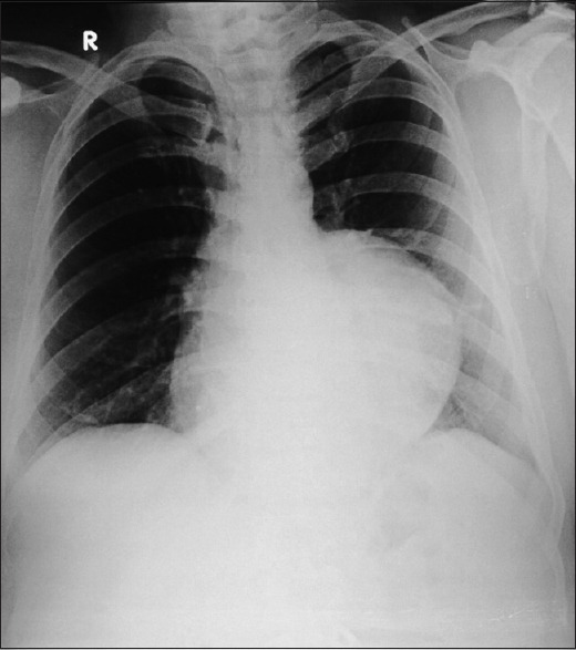
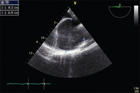
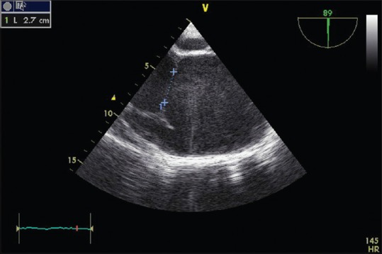
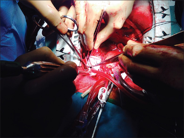
Free wall rupture of the left ventricle (LV) is a rare but life-threatening complication of acute myocardial infaction. Very rarely such rupture may be contained by the adhering pericardium creating a pseudoaneurysm. This condition warrants for an emergency surgery. Left ventricular aneurysm is the discrete thinning of the ventricular wall (<5 mm) with akinetic or dyskinetic wall motion causing an out-pouching of the ventricle. Given the propensity for pseudoaneurysms to rupture leading to cardiac tamponade, shock, and death, compared with a more benign natural history for true aneurysms, accurate diagnosis of these conditions is important. True aneurysm, usually, calls for an elective surgery. Clinically differentiating the two conditions remains a challenge. We report the case of a patient with LV pseudoaneurysm, initially diagnosed as true aneurysm at our institution. We have attempted to review the existing literature and discussed the characteristic findings of each entity.
Figures
References
-
- Pollak H, Nobis H, Mlczoch J. Frequency of left ventricular free wall rupture complicating acute myocardial infarction since the advent of thrombolysis. Am J Cardiol. 1994;74:184–6. - PubMed
-
- Brown SL, Gropler RJ, Harris KM. Distinguishing left ventricular aneurysm from pseudoaneurysm. A review of the literature. Chest. 1997;111:1403–9. - PubMed
-
- Dachman AH, Spindola-Franco H, Solomon N. Left ventricular pseudoaneurysm. Its recognition and significance. JAMA. 1981;246:1951–3. - PubMed
-
- Gueron M, Wanderman KL, Hirsch M, Borman J. Pseudoaneurysm of the left ventricle after myocardial infarction: A curable form of myocardial rupture. J Thorac Cardiovasc Surg. 1975;69:736–42. - PubMed
Publication types
MeSH terms
LinkOut - more resources
Full Text Sources
Other Literature Sources