Review
. 2016 Jan 6;6(1):6.
doi: 10.3390/biom6010006.

Tau Protein Hyperphosphorylation and Aggregation in Alzheimer's Disease and Other Tauopathies, and Possible Neuroprotective Strategies

Affiliations
Review

Tau Protein Hyperphosphorylation and Aggregation in Alzheimer's Disease and Other Tauopathies, and Possible Neuroprotective Strategies

Goran Šimić et al. Biomolecules. .

Abstract

Abnormal deposition of misprocessed and aggregated proteins is a common final pathway of most neurodegenerative diseases, including Alzheimer's disease (AD). AD is characterized by the extraneuronal deposition of the amyloid β (Aβ) protein in the form of plaques and the intraneuronal aggregation of the microtubule-associated protein tau in the form of filaments. Based on the biochemically diverse range of pathological tau proteins, a number of approaches have been proposed to develop new potential therapeutics. Here we discuss some of the most promising ones: inhibition of tau phosphorylation, proteolysis and aggregation, promotion of intra- and extracellular tau clearance, and stabilization of microtubules. We also emphasize the need to achieve a full understanding of the biological roles and post-translational modifications of normal tau, as well as the molecular events responsible for selective neuronal vulnerability to tau pathology and its propagation. It is concluded that answering key questions on the relationship between Aβ and tau pathology should lead to a better understanding of the nature of secondary tauopathies, especially AD, and open new therapeutic targets and strategies.

Keywords: Alzheimer’s disease; amyloid β; microtubules; neurofibrillary degeneration; neuropathology; phosphorylation; protein aggregation; protein oligomerization; tau protein; tauopathies.


Figures

Figure 1
Figure 1
Chromosomal location of the gene and protein structure for the microtubule-associated proteins tau, microtubule-associated protein 2 (MAP2) and MAP4. Tau exons 2, 3 and 10 are alternatively spliced, giving rise to six different mRNAs, translated in six different tau isoforms. Tau isoforms differ by the absence or presence of one or two 29 amino acid inserts encoded by exon 2 (yellow) and 3 (orange) in the N-terminal part, in combination with either three (R1, R3 and R4) or four (R1-R4) repeat regions in the C-terminal part. The R2 repeat is encoded by exon 10. The longest 2N4R adult tau isoform (2+3+10+) has 441 amino acids (aa), followed by 1N4R isoform of 412 aa (2+3−10+), 2N3R isoform of 410 aa (2+3+10−), 0N4R isoform of 383 aa (2−3−10+), 2N3R isoform of 381aa (2+3−10−) and the shortest 0N3R isoform of 352 aa (2−3−10−). The single neuron-specific promoter of MAPT gene has three binding sites for transcription factors and its activity increases with axon initiation and outgrowth. The shortest tau isoform is the only one expressed in the fetal brain (“fetal tau”), while expression of other isoforms begins postnatally (for a review, see [48]). The MAP2 and MAP4 have comparable repeat domain sequences in the C-terminus but differ from tau proteins by their longer N-terminal projection arms.
Figure 2
Figure 2
Primary sequence of amino acids and probable secondary structure of the longest tau isoform in the central nervous system. N1 and N2 denote the sequences encoded by exons 2 and 3, respectively. R1 through R4 are microtubule-binding domains encoded by exons 9–12, respectively. Domains with β-sheet structure and α-helical content are shown in yellow and red, respectively.
Figure 3
Figure 3
Putative phosphorylation sites on tau protein and epitopes specific for major tau antibodies. Red color denotes amino acids phosphorylated in AD brain, green in both AD and normal brain, blue in normal brain, while black color means that those phosphorylation sites have not been fully characterized yet. Tau antibodies specific for phospho-tau epitopes are given in purple, while pink color denotes antibodies specific for non-phosphorylated tau epitopes: Alz-50 (aa 2–10, aa 312−342), 43D (aa 1–100), 77E9 (aa 185–195), 39E10 (aa 189–195), Tau-5 (aa 210–230), 5C7 (aa 267–278), Tau-1 (aa 195, 198, 199 and 202), 77G7 (aa 270–375), Tau-46 (aa 404–441), TauC-3 (tau cleaved on aa 421). Red—in the AD brain; Green—in both the AD and the normal brain; Blue—in the normal brain; Black—phosphorylation sites that have not been fully characterized yet; Purple—tau antibodies specific for phospho-tau epitopes; Pink—tau antibodies specific for unphosphorylated tau epitopes.
Figure 4
Figure 4
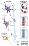
The sequence of cytoskeletal changes due to the pathology of tau protein divided into three stages: pre-tangle (pre-NFT) stage, and intraneuronal and extraneuronal stages. See text and Figure 5 for details.
Figure 5
Figure 5
Diagram showing sites for potential cleavage of tau protein. The sequential cleavage of the tau protein leads to the formation of the tau protein fragment from the microtubule-binding repeat region (see text and Figure 4). Tau cleavage is more likely to take place while protein is unbound to microtubules, either aggregated to itself or associated with proteins other than tubulin. PSA = puromycin-sensitive aminopeptidase.
Figure 6
Figure 6
Schematic representation of three different ways of anterograde spreading of tau aggregates by endocytosis, macropinocytosis, and exosomes.
Figure 7
Figure 7
Diagram showing potential neuroprotective strategies to reduce tau aggregates. See text for details.
Figure 8
Figure 8
Diagram of tau aggregation inhibitor LMTX (leucomethylthioninium with a suitable counterion), and its presumed mode of action (inhibition of tau aggregation).

References

    1. Bielschowsky M. Die Silberimprägnation der Achsenzylinder. Neurol. Zentralb. (Leipzig) 1902;13:579–584. (In German)
    1. Alzheimer A. Uber eine eigenartige Erkrankung der Hirnrinde. Allg Zeits Psychiatry Psych. Med. 1907;64:146–148. (In German)
    1. Jucker M., Beyreuther K., Haass C., Nitsch R., Christen Y. Alzheimer: 100 Years and Beyond. Springer; Berlin, Germany: 2006.
    1. Kidd M. Paired helical filaments in electron microscopy of Alzheimer’s disease. Nature. 1963;197:192–193. doi: 10.1038/197192b0. - DOI - PubMed
    1. Terry R.D. The fine structure of neurofibrillary tangles in Alzheimer’s disease. J. Neuropathol. Exp. Neurol. 1963;22:629–642. doi: 10.1097/00005072-196310000-00005. - DOI - PubMed

Publication types

MeSH terms