Review
. 2016 Jan 6;6(1):7.
doi: 10.3390/biom6010007.

Characteristics of Tau and Its Ligands in PET Imaging

Affiliations
Review

Characteristics of Tau and Its Ligands in PET Imaging

Ryuichi Harada et al. Biomolecules. .

Abstract

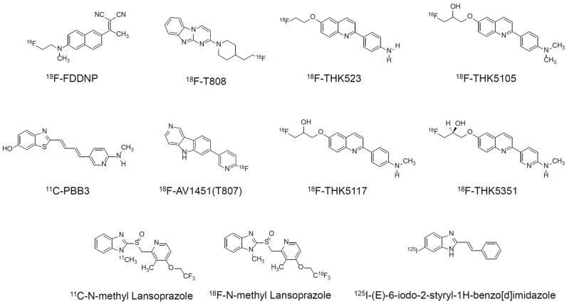
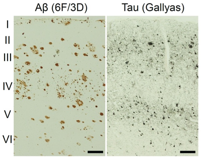
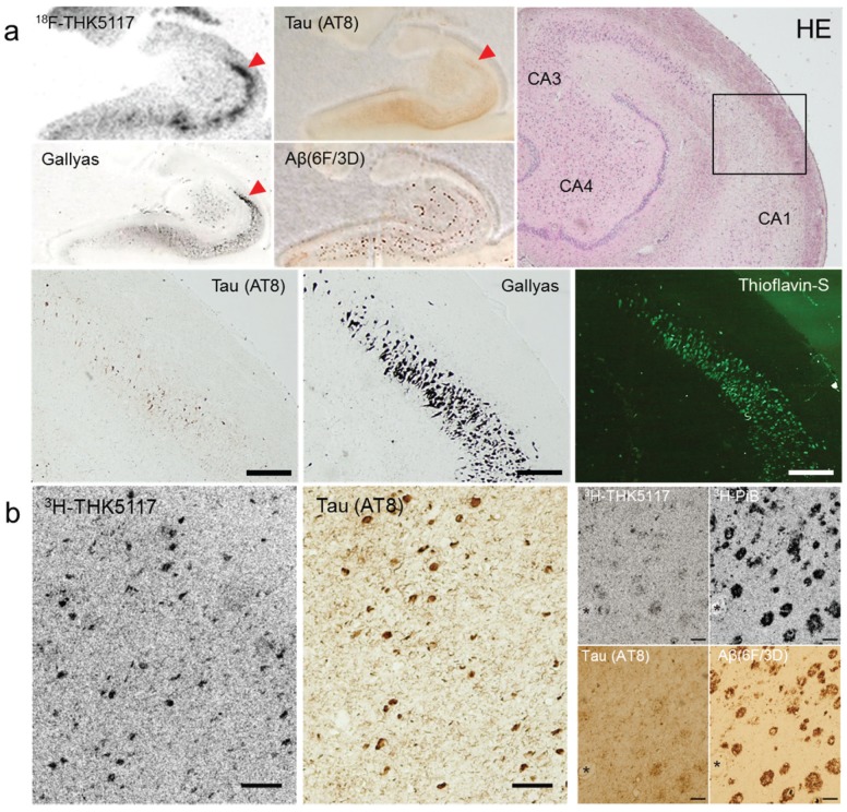
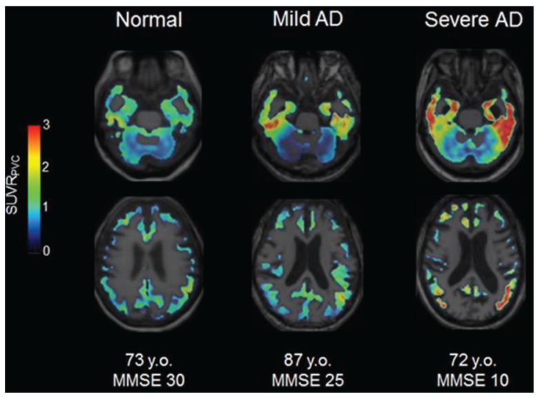
Tau deposition is one of the neuropathological hallmarks in Alzheimer's disease as well as in other neurodegenerative disorders called tauopathies. Recent efforts to develop selective tau radiopharmaceuticals have allowed the visualization of tau deposits in vivo. In vivo tau imaging allows the assessment of the regional distribution of tau deposits in a single human subject over time for determining the pathophysiology of tau accumulation in aging and neurodegenerative conditions as well as for application in drug discovery of anti-dementia drugs as surrogate markers. However, tau deposits show complicated characteristics because of different isoform composition, histopathology, and ultrastructure in various neurodegenerative conditions. In addition, since tau radiopharmaceuticals possess different chemotype classes, they may show different binding characteristics with heterogeneous tau deposits. In this review, we describe the characteristics of tau deposits and their ligands that have β-sheet binding properties, and the status of tau imaging in clinical studies.

Keywords: Alzheimer’s disease; positron emission tomography; radiotracer; tau deposits.


Figures

Figure 1
Figure 1
Chemical structures of different chemotype classes of tau ligands.
Figure 2
Figure 2
Different distribution of amyloid pathology and neurofibrillary pathology stained with anti-Aβ (6F/3D) antibody and Gallyas silver staining, respectively. Scale bar: 200 μm.
Figure 3
Figure 3
(a) In vitro autoradiography of 18F-THK5117 and tau/amyloid immunohistochemistry, H&E staining, Gallyas silver staining, and thioflavin-S fluorescence staining in the hippocampus of AD brain sections. Scale bars: 400 μm. (b) Microscopic observation of 3H-THK5117 and 3H-PiB labeled sections after photo emulsion treatment and tau/amyloid immunohistochemistry in adjacent sections. (asterisks indicate same blood vessel, scale bars 100 μm).
Figure 4
Figure 4
Fluorescence staining of THK5117 in various tauopathies such as tangle predominant senile dementia (TPSD), argyrophilic grain disease (AGD), Pick’s disease, corticobasal degeneration (CBD), and progressive supranuclear palsy (PSP). Scale bar: 20 μm. Arrows indicate the same tau deposits for each brain sample.
Figure 5
Figure 5
18F-THK5117 PET imaging in a normal elderly subject (Mini Mental State Examination (MMSE) 30, 73 y.o.), patient with mild AD (MMSE 25, 87 y.o.) and patient with severe AD (MMSE 10, 72 y.o.).

References

    1. Stein T.D., Alvarez V.E., McKee A.C. Chronic traumatic encephalopathy: A spectrum of neuropathological changes following repetitive brain trauma in athletes and military personnel. Alzheimer’s Res. Ther. 2014 doi: 10.1186/alzrt234. - DOI - PMC - PubMed
    1. Braak H., Braak E. Neuropathological stageing of Alzheimer-related changes. Acta Neuropathol. 1991;82:239–259. doi: 10.1007/BF00308809. - DOI - PubMed
    1. Delacourte A., David J.P., Sergeant N., Buee L., Wattez A., Vermersch P., Ghozali F., Fallet-Bianco C., Pasquier F., Lebert F., et al. The biochemical pathway of neurofibrillary degeneration in aging and Alzheimer’s disease. Neurology. 1999;52:1158–1165. doi: 10.1212/WNL.52.6.1158. - DOI - PubMed
    1. Arriagada P.V., Growdon J.H., Hedley-Whyte E.T., Hyman B.T. Neurofibrillary tangles but not senile plaques parallel duration and severity of Alzheimer’s disease. Neurology. 1992;42:631–639. doi: 10.1212/WNL.42.3.631. - DOI - PubMed
    1. Bierer L.M., Hof P.R., Purohit D.P., Carlin L., Schmeidler J., Davis K.L., Perl D.P. Neocortical neurofibrillary tangles correlate with dementia severity in Alzheimer’s disease. Arch. Neurol. 1995;52:81–88. doi: 10.1001/archneur.1995.00540250089017. - DOI - PubMed

Publication types