. 2016 Aug 11;35(32):4179-87.
doi: 10.1038/onc.2015.483. Epub 2016 Jan 11.

CFTR is a tumor suppressor gene in murine and human intestinal cancer

Affiliations

CFTR is a tumor suppressor gene in murine and human intestinal cancer

B L N Than et al. Oncogene. .

Erratum in

  • CFTR is a tumor suppressor gene in murine and human intestinal cancer.
    Than BLN, Linnekamp JF, Starr TK, Largaespada DA, Rod A, Zhang Y, Bruner V, Abrahante J, Schumann A, Luczak T, Walter J, Niemczyk A, O'Sullivan MG, Medema JP, Fijneman RJA, Meijer GA, Van den Broek E, Hodges CA, Scott PM, Vermeulen L, Cormier RT. Than BLN, et al. Oncogene. 2017 Jun 15;36(24):3504. doi: 10.1038/onc.2017.3. Epub 2017 Feb 13. Oncogene. 2017. PMID: 28192405

Abstract

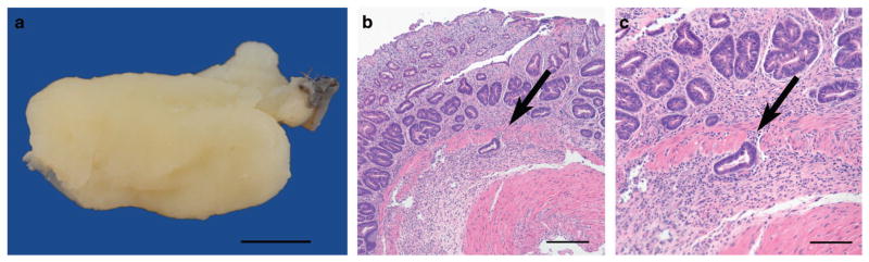
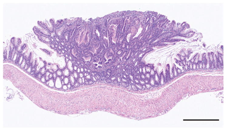
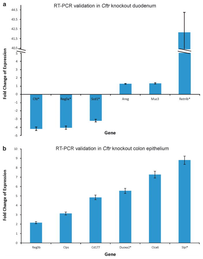
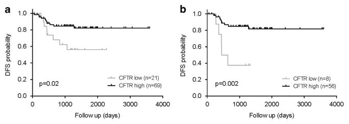
CFTR, the cystic fibrosis (CF) gene, encodes for the CFTR protein that plays an essential role in anion regulation and tissue homeostasis of various epithelia. In the gastrointestinal (GI) tract CFTR promotes chloride and bicarbonate secretion, playing an essential role in ion and acid-base homeostasis. Cftr has been identified as a candidate driver gene for colorectal cancer (CRC) in several Sleeping Beauty DNA transposon-based forward genetic screens in mice. Further, recent epidemiological and clinical studies indicate that CF patients are at high risk for developing tumors in the colon. To investigate the effects of CFTR dysregulation on GI cancer, we generated Apc(Min) mice that carried an intestinal-specific knockout of Cftr. Our results indicate that Cftr is a tumor suppressor gene in the intestinal tract as Cftr mutant mice developed significantly more tumors in the colon and the entire small intestine. In Apc(+/+) mice aged to ~1 year, Cftr deficiency alone caused the development of intestinal tumors in >60% of mice. Colon organoid formation was significantly increased in organoids created from Cftr mutant mice compared with wild-type controls, suggesting a potential role of Cftr in regulating the intestinal stem cell compartment. Microarray data from the Cftr-deficient colon and the small intestine identified dysregulated genes that belong to groups of immune response, ion channel, intestinal stem cell and other growth signaling regulators. These associated clusters of genes were confirmed by pathway analysis using Ingenuity Pathway Analysis and gene set enrichment analysis (GSEA). We also conducted RNA Seq analysis of tumors from Apc(+/+) Cftr knockout mice and identified sets of genes dysregulated in tumors including altered Wnt β-catenin target genes. Finally we analyzed expression of CFTR in early stage human CRC patients stratified by risk of recurrence and found that loss of expression of CFTR was significantly associated with poor disease-free survival.


Conflict of interest statement

CONFLICT OF INTEREST

The authors declare no conflict of interest.

Figures

Figure 1
Figure 1
Tumor in the rectum of ApcMin Cftrfl/fl-Villin-Cre mouse. Diffuse mucosal thickening in prolapsed rectal mucosa is shown in (a) (bar =5 mm). There is adenomatous hyperplasia with disorganization, irregular tortuosity and branching of crypts with focal invasion into the submucosa (arrow) (panel b, bar = 200 μm); focal invasion of the submucosa by a proliferating tubule is indicated by an arrow (panel c, bar =100 μm).
Figure 2
Figure 2
Tumor in the small intestine of Apc+/+ Cftrfl/fl-Villin-Cre mouse. A polypoid adenoma (bar = 500 μm).
Figure 3
Figure 3
(a) Quantitative reverse transcriptase PCR (qRT–PCR) gene expression analysis of mouse normal small intestine of Apc+/+ Cftrfl/fl-Villin-Cre mice. Each gene sample was run in triplicate and gene expression was normalized to the expression of 18S. Data are presented as the mean fold change ±s.d. Each bar represents the mean and s.e. of multiple experiments that measured fold differences in the mRNA expression in proximal small intestine tissue isolated from adult (~100 days) littermate and gender matched pairs of Apc+/+ Cftrfl/fl-Villin-Cre and Cftr+/+ mice. mRNAs were isolated from 1 cm sections of the proximal small intestine from the same region for all mice. Villi were removed from the tissue prior to processing. Four replicates of each assay were performed for each matched pair of mRNAs and these sets of assays were repeated at least two times for each pair of mRNAs. At least two matched pairs of mRNAs were tested for each gene with most genes tested in at least three matched pairs of mRNAs. To be included in this figure genes met the flowing criteria: (1) the mean fold difference was at least 1.5; and (2) each gene showed a change in gene expression in the same direction in each matched pair of mRNAs. In all cases the direction of changes in gene expression confirmed microarray data. *P<0.05. (b) qRT–PCR gene expression analysis of mouse normal colon of Apc+/+ Cftrfl/fl-Villin-Cre mice. Samples were analyzed in triplicate and normalized to 18S ribosomal RNA. Data are presented as the mean fold change ± s.d. Each bar represents the mean and s.e. of multiple experiments that measured fold differences in the mRNA expression of whole colon tissue isolated from adult (~100 days), littermate and gender matched pairs of Apc+/+ Cftrfl/fl-Villin-Cre and Cftr+/+ mice. RNA was isolated from 1 cm sections from the same region of distal colon. Four replicates of each assay were performed for each matched pair of mRNAs and these sets of assays were repeated at least two times. At least two matched pairs of mRNAs were tested for each gene with most genes tested in at least three matched pairs of mRNAs. To be included in this figure genes met the flowing criteria: (1) the mean fold difference was at least 1.5; and (2) each gene showed a change in gene expression in the same direction in each matched pair of mRNAs. In all cases the direction in changes in gene expression confirmed microarray data. *P<0.05.
Figure 4
Figure 4
Kaplan–Meier estimate of disease-free survival (DFS) in the AMC-AJCCII-90 set between CFTR low and CFTR high-expressing tumors. Disease-free survival within the AMC-AJCCII-90 set stratified by CFTR expression of the tumor in the complete set (a) and in the MSS subset (b). The 3-year disease-free survival for the complete set (a) was 85% in the CFTR high group and 56% in the CFTR low group. In the MSS set (b) the 3-year disease-free survival was 85 and 38% in the CFTR high group and CFTR low group, respectively. P-values are based on log-rank test.

References

    1. De Lisle RC, Borowitz D. The cystic fibrosis intestine. Cold Spring Harb Perspect Med. 2013;3:a009753. - PMC - PubMed
    1. Riordan JR, Rommens JM, Kerem B, Alon N, Rozmahel R, Grzelczak Z, et al. Identification of the cystic fibrosis gene: cloning and characterization of complementary DNA. Science. 1989;245:1066–1073. - PubMed
    1. Pedersen SF, Stock C. Ion channels and transporters in cancer: pathophysiology, regulation, and clinical potential. Cancer Res. 2013;73:1658–1661. - PubMed
    1. Neglia JP, FitzSimmons SC, Maisonneuve P, Schöni MH, Schöni-Affolter F, Corey M, et al. The risk of cancer among patients with cystic fibrosis. Cystic Fibrosis and Cancer Study Group. N Engl J Med. 1995;332:494–499. - PubMed
    1. McWilliams R, Highsmith WE, Rabe KG, de Andrade M, Tordsen LA, Holtegaard LM, et al. Cystic fibrosis transmembrane regulator gene carrier status is a risk factor for young onset pancreatic adenocarcinoma. Gut. 2005;54:1661–1662. - PMC - PubMed

Publication types

Substances