

The CaSm (LSm1) oncogene promotes transformation, chemoresistance and metastasis of pancreatic cancer cells
- PMID: 26751936
- PMCID: PMC4728675
- DOI: 10.1038/oncsis.2015.45
The CaSm (LSm1) oncogene promotes transformation, chemoresistance and metastasis of pancreatic cancer cells
Abstract
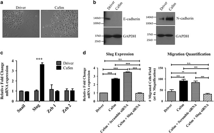
The cancer-associated Sm-like (CaSm) oncogene is overexpressed in 87% of human pancreatic tumor samples and CaSm knockdown has demonstrated therapeutic efficacy in murine models of pancreatic cancer. Evidence indicates that CaSm modulates messenger RNA degradation; however, its target genes and the mechanisms by which CaSm promotes pancreatic cancer remain largely unknown. Here, we demonstrate that the CaSm overexpression alters several hallmarks of cancer-including transformation, proliferation, chemoresistance and metastasis. Doxycycline-induced CaSm expression enhanced proliferation and both anchorage-dependent and -independent growth of the human Panc-1 cells in vitro. CaSm induction decreased gemcitabine-induced cytotoxicity and altered the expression of apoptotic regulation genes, including Bad, E2F1 and Bcl-XL. CaSm-overexpressing Panc-1 cells were twofold more migratory and fourfold more invasive than the driver controls and demonstrated characteristics of epithelial-to-mesenchymal transition such as morphological changes and decreased E-cadherin expression. CaSm induction resulted in changes in RNA expression of metastasis-associated genes such as MMP1, SerpinB5, uPAR and Slug. Using a murine model of metastatic pancreatic cancer, injection of CaSm-induced Panc-1 cells resulted in a higher abundance of hepatic metastatic lesions. Overall, CaSm overexpression contributed to a more aggressive cancer phenotype in Panc-1 cells, further supporting the use of CaSm as a therapeutic target against pancreatic cancer.
Figures


Similar articles
-
Establishing a murine pancreatic cancer CaSm model: up-regulation of CaSm is required for the transformed phenotype of murine pancreatic adenocarcinoma.Mol Ther. 2005 Mar;11(3):363-72. doi: 10.1016/j.ymthe.2004.09.023. Mol Ther. 2005. PMID: 15727932
-
CaSm/gemcitabine chemo-gene therapy leads to prolonged survival in a murine model of pancreatic cancer.Surgery. 2001 Aug;130(2):280-8. doi: 10.1067/msy.2001.115899. Surgery. 2001. PMID: 11490361
-
CaSm-mediated cellular transformation is associated with altered gene expression and messenger RNA stability.Cancer Res. 2005 Jul 15;65(14):6228-36. doi: 10.1158/0008-5472.CAN-05-0650. Cancer Res. 2005. PMID: 16024624
-
CaSm antisense gene therapy: a novel approach for the treatment of pancreatic cancer.Anticancer Res. 2003 May-Jun;23(3A):2007-13. Anticancer Res. 2003. PMID: 12894573 Review.
-
CaSm (LSm-1) overexpression in lung cancer and mesothelioma is required for transformed phenotypes.Am J Respir Cell Mol Biol. 2008 Jun;38(6):671-8. doi: 10.1165/rcmb.2007-0205OC. Epub 2008 Jan 24. Am J Respir Cell Mol Biol. 2008. PMID: 18218995 Free PMC article.
Cited by
-
m7G regulator-mediated molecular subtypes and tumor microenvironment in kidney renal clear cell carcinoma.Front Pharmacol. 2022 Sep 6;13:900006. doi: 10.3389/fphar.2022.900006. eCollection 2022. Front Pharmacol. 2022. PMID: 36147333 Free PMC article.
-
Prognostic value and potential molecular mechanism of the like-Sm gene family in early-stage pancreatic ductal adenocarcinoma.Transl Cancer Res. 2021 Apr;10(4):1744-1760. doi: 10.21037/tcr-20-3056. Transl Cancer Res. 2021. PMID: 35116499 Free PMC article.
-
Processing body (P-body) and its mediators in cancer.Mol Cell Biochem. 2022 Apr;477(4):1217-1238. doi: 10.1007/s11010-022-04359-7. Epub 2022 Jan 28. Mol Cell Biochem. 2022. PMID: 35089528 Review.
-
Sm-like 5 knockdown inhibits proliferation and promotes apoptosis of colon cancer cells by upregulating p53, CDKN1A and TNFRSF10B.World J Gastrointest Oncol. 2024 Jun 15;16(6):2716-2726. doi: 10.4251/wjgo.v16.i6.2716. World J Gastrointest Oncol. 2024. PMID: 38994171 Free PMC article.
-
Correlation between LSM1 Expression and Clinical Outcomes in Glioblastoma and Functional Mechanisms.Int J Genomics. 2023 Nov 2;2023:1543620. doi: 10.1155/2023/1543620. eCollection 2023. Int J Genomics. 2023. PMID: 37954131 Free PMC article.
References
-
- 1National Cancer Institute Pancreatic Cancer Treatment PDQ. Available from http://www.cancer.gov/cancertopics/pdq/treatment/pancreatic/healthprofes....
-
- 2Burris HA 3rd, Moore MJ, Andersen J, Green MR, Rothenberg ML, Modiano MR et al. Improvements in survival and clinical benefit with gemcitabine as first-line therapy for patients with advanced pancreas cancer: a randomized trial. J Clin Oncol 1997; 15: 2403–2413. - PubMed
-
- 3Thota R, Pauff JM, Berlin JD. Treatment of metastatic pancreatic adenocarcinoma: a review. Oncology (Williston Park) 2014; 28: 70–74. - PubMed
-
- 4Arshad A, Al-Leswas D, Al-Taan O, Stephenson J, Metcalfe M, Steward WP et al. Pooled survival and response data from phase III randomized controlled trials for gemcitabine-based regimes in the treatment of advanced pancreatic cancer. Am J Clin Oncol 2011; 36: 411–414. - PubMed
-
- 5Shi S, Yao W, Xu J, Long J, Liu C, Yu X. Combinational therapy: new hope for pancreatic cancer? Cancer Lett 2012; 317: 127–135. - PubMed
Grants and funding
LinkOut - more resources
Full Text Sources
Other Literature Sources
Molecular Biology Databases
Research Materials
Miscellaneous
