Curcumin Suppresses Proliferation and Migration of MDA-MB-231 Breast Cancer Cells through Autophagy-Dependent Akt Degradation
- PMID: 26752181
- PMCID: PMC4708990
- DOI: 10.1371/journal.pone.0146553
Curcumin Suppresses Proliferation and Migration of MDA-MB-231 Breast Cancer Cells through Autophagy-Dependent Akt Degradation
Retraction in
-
Retraction: Curcumin Suppresses Proliferation and Migration of MDA-MB-231 Breast Cancer Cells through Autophagy-Dependent Akt Degradation.PLoS One. 2023 Mar 15;18(3):e0283354. doi: 10.1371/journal.pone.0283354. eCollection 2023. PLoS One. 2023. PMID: 36920966 Free PMC article. No abstract available.
Abstract
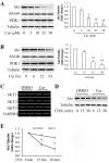
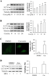
Previous studies have evidenced that the anticancer potential of curcumin (diferuloylmethane), a main yellow bioactive compound from plant turmeric was mediated by interfering with PI3K/Akt signaling. However, the underlying molecular mechanism is still poorly understood. This study experimentally revealed that curcumin treatment reduced Akt protein expression in a dose- and time-dependent manner in MDA-MB-231 breast cancer cells, along with an activation of autophagy and suppression of ubiquitin-proteasome system (UPS) function. The curcumin-reduced Akt expression, cell proliferation, and migration were prevented by genetic and pharmacological inhibition of autophagy but not by UPS inhibition. Additionally, inactivation of AMPK by its specific inhibitor compound C or by target shRNA-mediated silencing attenuated curcumin-activated autophagy. Thus, these results indicate that curcumin-stimulated AMPK activity induces activation of the autophagy-lysosomal protein degradation pathway leading to Akt degradation and the subsequent suppression of proliferation and migration in breast cancer cell.
Conflict of interest statement
Figures


Similar articles
-
Cycle arrest and apoptosis in MDA-MB-231/Her2 cells induced by curcumin.Eur J Pharmacol. 2012 Sep 5;690(1-3):22-30. doi: 10.1016/j.ejphar.2012.05.036. Epub 2012 Jun 15. Eur J Pharmacol. 2012. PMID: 22705896
-
Antitumor activity of curcumin by modulation of apoptosis and autophagy in human lung cancer A549 cells through inhibiting PI3K/Akt/mTOR pathway.Oncol Rep. 2018 Mar;39(3):1523-1531. doi: 10.3892/or.2018.6188. Epub 2018 Jan 4. Oncol Rep. 2018. PMID: 29328421
-
Curcumin prevented human autocrine growth hormone (GH) signaling mediated NF-κB activation and miR-183-96-182 cluster stimulated epithelial mesenchymal transition in T47D breast cancer cells.Mol Biol Rep. 2019 Feb;46(1):355-369. doi: 10.1007/s11033-018-4479-y. Epub 2018 Nov 23. Mol Biol Rep. 2019. PMID: 30467667
-
Targeting the Akt signaling pathway: Exploiting curcumin's anticancer potential.Pathol Res Pract. 2024 Sep;261:155479. doi: 10.1016/j.prp.2024.155479. Epub 2024 Jul 20. Pathol Res Pract. 2024. PMID: 39068859 Review.
-
The Function of Autophagy in the Initiation, and Development of Breast Cancer.Curr Med Chem. 2024;31(20):2974-2990. doi: 10.2174/0929867330666230503145319. Curr Med Chem. 2024. PMID: 37138421 Review.
Cited by
-
4-Methoxydalbergione Elicits Anticancer Effects by Upregulation of GADD45G in Human Liver Cancer Cells.J Healthc Eng. 2023 Feb 1;2023:6710880. doi: 10.1155/2023/6710880. eCollection 2023. J Healthc Eng. 2023. PMID: 36776954 Free PMC article.
-
The "Yin and Yang" of Natural Compounds in Anticancer Therapy of Triple-Negative Breast Cancers.Cancers (Basel). 2018 Sep 21;10(10):346. doi: 10.3390/cancers10100346. Cancers (Basel). 2018. PMID: 30248941 Free PMC article. Review.
-
Curcumin Induces Autophagy, Apoptosis, and Cell Cycle Arrest in Human Pancreatic Cancer Cells.Evid Based Complement Alternat Med. 2017;2017:5787218. doi: 10.1155/2017/5787218. Epub 2017 Sep 10. Evid Based Complement Alternat Med. 2017. PMID: 29081818 Free PMC article.
-
Sinapic Acid Attenuates Chronic DSS-Induced Intestinal Fibrosis in C57BL/6J Mice by Modulating NLRP3 Inflammasome Activation and the Autophagy Pathway.ACS Omega. 2023 Dec 26;9(1):1230-1241. doi: 10.1021/acsomega.3c07474. eCollection 2024 Jan 9. ACS Omega. 2023. PMID: 38222654 Free PMC article.
-
ACNPD: The Database for Elucidating the Relationships Between Natural Products, Compounds, Molecular Mechanisms, and Cancer Types.Front Pharmacol. 2021 Aug 23;12:746067. doi: 10.3389/fphar.2021.746067. eCollection 2021. Front Pharmacol. 2021. PMID: 34497528 Free PMC article.
References
Publication types
MeSH terms
Substances
LinkOut - more resources
Full Text Sources
Other Literature Sources
Medical
Miscellaneous

