

Differential Expression of Proteins Associated with the Hair Follicle Cycle - Proteomics and Bioinformatics Analyses
- PMID: 26752403
- PMCID: PMC4709225
- DOI: 10.1371/journal.pone.0146791
Differential Expression of Proteins Associated with the Hair Follicle Cycle - Proteomics and Bioinformatics Analyses
Abstract
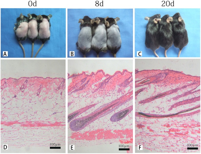
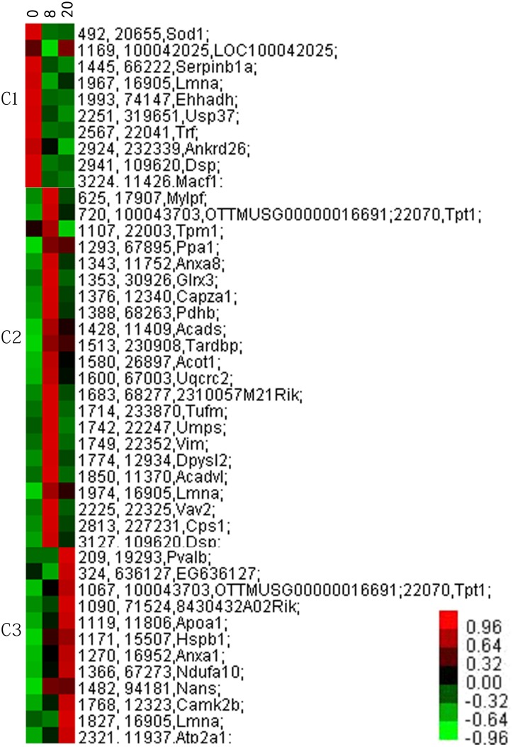
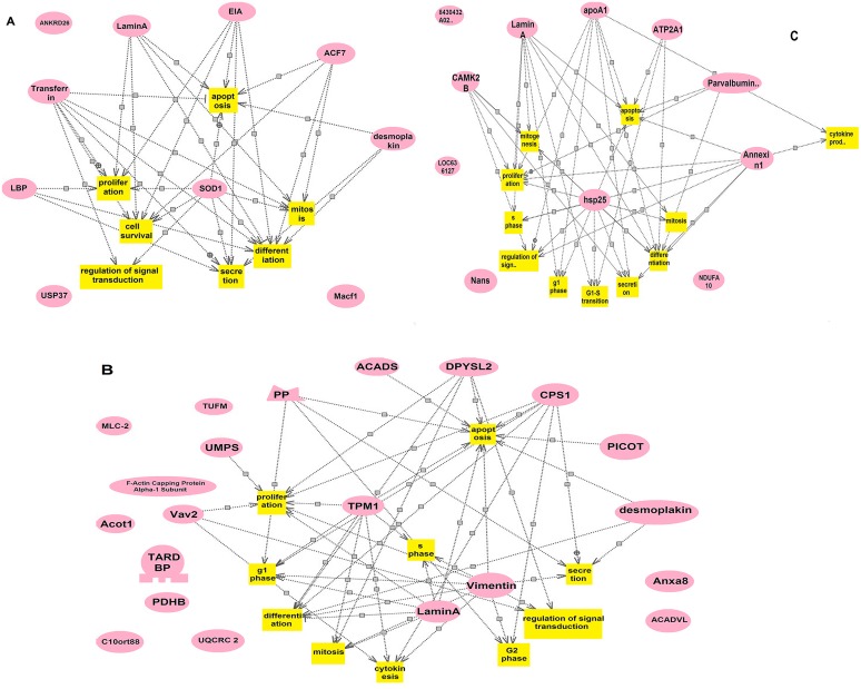
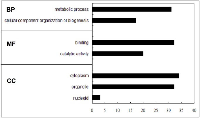
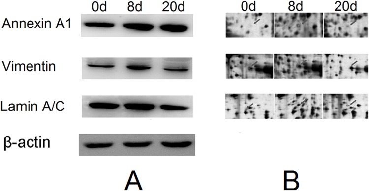
Hair follicle cycling can be divided into the following three stages: anagen, catagen, and telogen. The molecular signals that orchestrate the follicular transition between phases are still unknown. To better understand the detailed protein networks controlling this process, proteomics and bioinformatics analyses were performed to construct comparative protein profiles of mouse skin at specific time points (0, 8, and 20 days). Ninety-five differentially expressed protein spots were identified by MALDI-TOF/TOF as 44 proteins, which were found to change during hair follicle cycle transition. Proteomics analysis revealed that these changes in protein expression are involved in Ca2+-regulated biological processes, migration, and regulation of signal transduction, among other processes. Subsequently, three proteins were selected to validate the reliability of expression patterns using western blotting. Cluster analysis revealed three expression patterns, and each pattern correlated with specific cell processes that occur during the hair cycle. Furthermore, bioinformatics analysis indicated that the differentially expressed proteins impacted multiple biological networks, after which detailed functional analyses were performed. Taken together, the above data may provide insight into the three stages of mouse hair follicle morphogenesis and provide a solid basis for potential therapeutic molecular targets for this hair disease.
Conflict of interest statement
Figures
Similar articles
-
The up-regulation of 14-3-3 proteins in Smad4 deficient epidermis and hair follicles at catagen.Proteomics. 2008 Jun;8(11):2230-43. doi: 10.1002/pmic.200700760. Proteomics. 2008. PMID: 18446800
-
Expression of decorin throughout the murine hair follicle cycle: hair cycle dependence and anagen phase prolongation.Exp Dermatol. 2014 Jul;23(7):486-91. doi: 10.1111/exd.12441. Exp Dermatol. 2014. PMID: 24816226
-
Identification of proteins involved in aggregation of human dermal papilla cells by proteomics.J Dermatol Sci. 2007 Dec;48(3):189-97. doi: 10.1016/j.jdermsci.2007.06.013. Epub 2007 Sep 17. J Dermatol Sci. 2007. PMID: 17875385
-
Emerging Diagnostic and Therapeutic Potentials of Human Hair Proteomics.Proteomics Clin Appl. 2018 Mar;12(2). doi: 10.1002/prca.201700048. Epub 2017 Oct 23. Proteomics Clin Appl. 2018. PMID: 28960873 Review.
-
The secret life of the hair follicle.Trends Genet. 1992 Feb;8(2):55-61. doi: 10.1016/0168-9525(92)90350-d. Trends Genet. 1992. PMID: 1566372 Review.
Cited by
-
iTRAQ-based quantitative proteomics revealing the therapeutic mechanism of a medicinal and edible formula YH0618 in reducing doxorubicin-induced alopecia by targeting keratins and TGF-β/Smad3 pathway.Heliyon. 2024 Jun 18;10(12):e33051. doi: 10.1016/j.heliyon.2024.e33051. eCollection 2024 Jun 30. Heliyon. 2024. PMID: 39021977 Free PMC article.
-
Effect of captopril on radiation-induced TGF-β1 secretion in EA.Hy926 human umbilical vein endothelial cells.Oncotarget. 2017 Mar 28;8(13):20842-20850. doi: 10.18632/oncotarget.15356. Oncotarget. 2017. PMID: 28209920 Free PMC article.
-
Transcriptome sequencing reveals the expression profiles of lncRNAs and mRNAs in goat skin tissues with different types of wool coats.Sci Rep. 2025 May 30;15(1):18977. doi: 10.1038/s41598-025-01187-9. Sci Rep. 2025. PMID: 40447626 Free PMC article.
-
Differential proteomics of lesional vs. non-lesional biopsies revealed non-immune mechanisms of alopecia areata.Sci Rep. 2018 Jan 11;8(1):521. doi: 10.1038/s41598-017-18282-1. Sci Rep. 2018. PMID: 29323127 Free PMC article.
-
Cutaneous transcriptome analysis in NIH hairless mice.PLoS One. 2017 Aug 7;12(8):e0182463. doi: 10.1371/journal.pone.0182463. eCollection 2017. PLoS One. 2017. PMID: 28787439 Free PMC article.
References
-
- Paus R, Muller-Rover S, Van Der Veen C, Maurer M, Eichmuller S, Ling G, et al. A comprehensive guide for the recognition and classification of distinct stages of hair follicle morphogenesis. J Invest Dermatol. 1999. 113(4): 523–32. - PubMed
-
- Tomita Y, Akiyama M, Shimizu H. PDGF isoforms induce and maintain anagen phase of murine hair follicles. J Dermatol Sci. 2006. 43(2): 105–15. - PubMed
Publication types
MeSH terms
Substances
LinkOut - more resources
Full Text Sources
Other Literature Sources
Molecular Biology Databases
Miscellaneous
