. 2016 Feb;126(2):745-61.
doi: 10.1172/JCI81950. Epub 2016 Jan 11.

CXCL13 drives spinal astrocyte activation and neuropathic pain via CXCR5

CXCL13 drives spinal astrocyte activation and neuropathic pain via CXCR5

Bao-Chun Jiang et al. J Clin Invest. 2016 Feb.

Abstract

Recent studies have implicated chemokines in microglial activation and pathogenesis of neuropathic pain. C-X-C motif chemokine 13 (CXCL13) is a B lymphocyte chemoattractant that activates CXCR5. Using the spinal nerve ligation (SNL) model of neuropathic pain, we found that CXCL13 was persistently upregulated in spinal cord neurons after SNL, resulting in spinal astrocyte activation via CXCR5 in mice. shRNA-mediated inhibition of CXCL13 in the spinal cord persistently attenuated SNL-induced neuropathic pain. Interestingly, CXCL13 expression was suppressed by miR-186-5p, a microRNA that colocalized with CXCL13 and was downregulated after SNL. Spinal overexpression of miR-186-5p decreased CXCL13 expression, alleviating neuropathic pain. Furthermore, SNL induced CXCR5 expression in spinal astrocytes, and neuropathic pain was abrogated in Cxcr5-/- mice. CXCR5 expression induced by SNL was required for the SNL-induced activation of spinal astrocytes and microglia. Intrathecal injection of CXCL13 was sufficient to induce pain hypersensitivity and astrocyte activation via CXCR5 and ERK. Finally, intrathecal injection of CXCL13-activated astrocytes induced mechanical allodynia in naive mice. Collectively, our findings reveal a neuronal/astrocytic interaction in the spinal cord by which neuronally produced CXCL13 activates astrocytes via CXCR5 to facilitate neuropathic pain. Thus, miR-186-5p and CXCL13/CXCR5-mediated astrocyte signaling may be suitable therapeutic targets for neuropathic pain.


Figures

Figure 10
Figure 10. Schematic shows spinal neuronal-glial interactions in neuropathic pain after peripheral nerve injury.
(A) Neuronal-glial interactions revealed in previous studies. After nerve injury, CCL2 and CX3CL1 are released from the central terminals of primary afferent neurons (presynaptic sites) and spinal neurons (postsynaptic sites), which activate CCR2 and CX3CR1, respectively, on spinal microglia. CCL2 and CXCL1 can be released from astrocytes to act on spinal neurons via CCR2 and CXCR2, respectively. Astrocytes may also produce CCL7 to recognize CCR2 on microglia. Upon activation, microglia produce cytokines TNF-α and IL-18 to activate astrocytes via TNFR and IL-18R. Microglia also produce the neurotrophin brain-derived neurotrophic factor (BDNF) via P2X4 activation to enhance nociceptive synaptic transmission (63, 68). (B) Neuronal-glial interactions revealed in the present study. SNL increases glutamate release from presynaptic terminals to activate NMDAR on postsynaptic neurons, leading to decreased expression of miR-186-5p, and this decrease results in CXCL13 upregulation in spinal neurons. Upon release, CXCL13 acts on CXCR5 in astrocytes and activates the ERK signaling pathway, leading to the expression and release of astroglial mediators, such as CCL2 and CCL7, to maintain microglial activation. Astrocytes may also produce ATP to activate microglia via P2X4, P2X7, and P2Y12 receptors (47, 63, 69). These astrocytic mediators can also directly potentiate nociceptive transmission via pre- and postsynaptic modulation (10, 52). Together, this CXCL13-CXCR5–mediated neuronal-astrocytic interaction in the dorsal horn can enhance and prolong neuropathic pain states.
Figure 9
Figure 9. Injection (i.t.) of CXCL13-treated astrocytes induces mechanical allodynia.
(A) Experimental protocol showing the preparation of astrocytes for i.t. injection. The differentiated astrocytes from WT or Cxcr5–/– mice were incubated with PBS or CXCL13, then collected, washed with PBS, and resuspended for i.t. injection into naive mice. (B) Injection (i.t.) of CXCL13-treated astrocytes from WT animals (n = 7) induced dramatic decrease in PWT, and this decrease was compromised after injection of Cxcr5-deficient astrocytes (n = 9). ##P < 0.01 vs. baseline, 1-way repeated measures ANOVA followed by Bonferroni’s test; *P < 0.05, 2-way repeated measures ANOVA followed by Bonferroni’s test. (C) CXCL13 induced rapid pERK expression in astrocyte cultures from WT, but not from Cxcr5 KO mice. **P < 0.01 vs. control, 1-way ANOVA followed by Bonferroni’s test. n = 3 for each treatment.
Figure 8
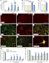
Figure 8. CXCL13 induces ERK-dependent astrocytic activation and pain hypersensitivity.
(A) Injection (i.t.) of CXCL13 increased pERK expression in the spinal cord in WT mice (A), but not in Cxcr5 KO mice (B). **P < 0.01, Student’s t test. n = 3 mice/group. (CF) Injection (i.t.) of CXCL13 increased pERK IR compared with i.t. vehicle (PBS) (C and D). pERK (D) was colocalized with GFAP (E and F) in the spinal cord 1 hour after injection. Scale bars: 50 μm. (G) Pretreatment with L-α-AA inhibited CXCL13-induced pERK expression in the spinal cord 1 hour after CXCL13 injection. *P < 0.05, vs. naive. #P < 0.05, vs. veh + CXCL13, Student’s t test. n = 4 mice/group. (H and I) Pretreatment with MEK inhibitor PD98059 blocked CXCL13-induced heat hyperalgesia (H) and mechanical allodynia (I). *P < 0.05; ***P < 0.001, 2-way repeated measures ANOVA followed by Bonferroni’s test. n = 6 mice/group. (J) PD98059 inhibited CXCL13-induced Gfap mRNA increase at 6 hours. **P < 0.01; ***P < 0.001, 1-way ANOVA followed by Bonferroni’s test. n = 3–5 mice/group. (K) Colocalization of pERK and GFAP in the spinal cord 10 days after SNL. Scale bars: 50 μm; 20 μm (insets). (L) pERK expression was increased in the spinal cord at 7 days and 28 days after SNL in WT mice, but not in Cxcr5 KO mice. *P < 0.05; ***P < 0.001, Student’s t test. n = 3 mice/group.
Figure 7
Figure 7. Injection (i.t.) of CXCL13 induces pain hypersensitivity and glial activation in the spinal cord.
(A) Injection (i.t.) of CXCL13 induced heat hyperalgesia in WT mice, but not in Cxcr5 KO mice. ***P < 0.001, WT vehicle (n = 7) vs. WT CXL13 (n = 7). ###P < 0.001, WT CXCL13 (n = 7) vs. Cxcr5–/– CXCL13 (n = 7), 2-way repeated measures ANOVA followed by Bonferroni’s test. (B) Injection (i.t.) of CXCL13 induced mechanical allodynia in WT mice, which was reduced in KO mice. ***P < 0.001, WT vehicle vs. WT CXL13. #P < 0.05; ##P < 0.01; ###P < 0.001, WT-CXCL13 vs. Cxcr5–/–-CXCL13, 2-way repeated measures ANOVA followed by Bonferroni’s test. (C) Injection (i.t.) of CXCL13 increased Gfap mRNA at 6 hours and Iba1 mRNA at 24 hours. **P < 0.01; ***P < 0.001 vs. control, 1-way ANOVA followed by Bonferroni’s test. n = 4–7 mice/group. (DH) Injection (i.t.) of CXCL13 increased GFAP IR in WT mice, but not in Cxcr5 KO mice 6 hours after injection. ***P < 0.001, Student’s t test. n = 4 mice/group. Scale bars: 100 μm. (I) Pretreatment with astroglial toxin L-α-AA blocked CXCL13-induced heat hyperalgesia. Pretreatment with microglial inhibitor minocycline attenuated heat hyperalgesia at 24 hours. *P < 0.05; ***P < 0.001, 2-way repeated measures ANOVA followed by Bonferroni’s test. n = 5 mice/group. (J) Pretreatment with L-α-AA inhibited CXCL13-induced mechanical allodynia. *P < 0.05, 2-way repeated measures ANOVA followed by Bonferroni’s test. n = 5–8 mice/group. (K) L-α-AA and minocycline decreased CXCL13-induced Iba1 mRNA upregulation in the spinal cord 24 hours after CXCL13 injection. *P < 0.05; **P < 0.01; ***P < 0.001, 1-way ANOVA followed by Bonferroni’s test. n = 3–6 mice/group. Veh, vehicle.
Figure 6
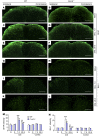
Figure 6. SNL-induced activation of glial cells in the spinal cord is inhibited in Cxcr5 KO mice.
(AF) GFAP staining in the spinal cord in naive (A and B), SNL at 7 days (C and D), and SNL at 28 days (E and F) of WT (A, C, and E) and Cxcr5 KO (B, D, and F) mice. (GL) IBA-1 staining in the spinal cord in naive (G and H), SNL at 7 days (I and J), and SNL at 28 days (K and L) of WT (G, I, and K) and Cxcr5 KO (H, J, and L) mice. Scale bars: 200 μm. (M and N) The intensity of GFAP (M) and IBA-1 (N) in the ipsilateral and contralateral spinal cord. **P < 0.01; ***P < 0.001, SNL vs. sham, Student’s t test. #P < 0.05; ##P < 0.01; ###P < 0.001, KO vs. WT, Student’s t test. N, naive. S, sham. n = 4 mice/group.
Figure 5
Figure 5. Cxcr5 is essential for SNL-induced neuropathic pain.
(AD) Acute pain thresholds and motor function are normal in Cxcr5 KO mice. Thermal sensitivity, measured by (A) tail immersion and (B) Hargreaves test, was comparable in WT and KO mice. Mechanical sensitivity assessed by von Frey test was indistinguishable in WT and KO mice (C). Motor function assessed by recording the falling latency in a Rotarod test was comparable in WT and KO mice (D). n = 11 mice/group (AC); n = 6 mice/group (D). (E and F) SNL-induced mechanical allodynia (E) and heat hyperalgesia (F) were largely reduced in Cxcr5 KO mice (n = 10) compared with WT mice (n = 8). ***P < 0.001, 2-way repeated measures ANOVA followed by Bonferroni’s test. (G and H) Intraspinal infusion of LV-Cxcr5 shRNA lentivirus in the spinal cord 10 days after SNL alleviated SNL-induced mechanical allodynia (G) and blocked heat hyperalgesia (H). *P < 0.05; **P < 0.01; ***P < 0.001, 2-way repeated measures ANOVA followed by Bonferroni’s test. n = 6 mice/group.
Figure 4
Figure 4. CXCR5 expression is increased in spinal astrocytes after SNL.
(A) Time course of Cxcr5 mRNA expression in the ipsilateral dorsal horn in naive (n = 6), sham-operated (n = 5), and SNL mice. SNL increased Cxcr5 expression at 1 day (n = 5), 3 days (n = 3), 10 days (n = 4), and 21 days (n = 4) compared with sham. *P < 0.05; ***P < 0.001, Student’s t test. (B) Western blotting shows the increase of CXCR5 protein in the spinal cord 10 days after SNL. *P < 0.05, Student’s t test. n = 3 mice/group. (CE) Representative images of CXCR5 immunofluorescence in the spinal cord from naive and SNL mice. CXCR5 IR was low in naive mice (C), increased in the ipsilateral dorsal horn of SNL mice (D), and unchanged in the contralateral dorsal horn of SNL mice (E). Scale bars: 100 μm. (FH) Double staining shows that CXCR5 is mainly colocalized with GFAP (F), rarely with NeuN (G), but not with OX-42 (H) in the dorsal horn of spinal cord 10 days after SNL. Scale bars: 50 μm; 10 μm (insets).
Figure 3
Figure 3. miR-186-5p inhibits neuropathic pain via suppressing CXCL13 expression in the spinal cord.
(A and B) Intraspinal infusion of mmu-pre-miR-186 expressing lentivirus 3 days after SNL alleviated SNL-induced mechanical allodynia (A) and heat hyperalgesia (B) at days 7, 10, and 14. *P < 0.05; ***P < 0.001, 2-way repeated measures ANOVA followed by Bonferroni’s test. n = 7 for LV-NC; n = 8 for SNL or LV-pre-mmu-miR-186 group. BL, baseline. (C and D) Injection (i.t.) of miR-186-5p mimic daily for 3 days (from day 8 to day 10 after SNL) attenuated SNL-induced mechanical allodynia (C) and heat hyperalgesia (D). **P < 0.01, ***P < 0.001, 2-way repeated measures ANOVA followed by Bonferroni’s test. n = 9 for NC and n = 10 for miR-186 mimic. (E) ELISA results show decreased CXCL13 levels in the spinal cord of miR-186-5p mimic–treated animals. *P < 0.05, Student’s t test. n = 6 mice/group. (F and G) Injection (i.t.) of miR-186-5p inhibitor induced mechanical allodynia (F) and heat hyperalgesia (G) in naive mice. *P < 0.05; **P < 0.01; ***P < 0.001, miR-186-5p inhibitor vs. NC, 2-way repeated measures ANOVA followed by Bonferroni’s test. n = 5 mice/group. (H) CXCL13 neutralizing antibody attenuated miR-186-5p inhibitor–induced mechanical allodynia. *P < 0.05, 2-way repeated measures ANOVA followed by Bonferroni’s test. n = 5 mice/group. (I) Injection (i.t.) of MK801 before SNL prevented SNL-induced downregulation of miR-186-5p in the spinal cord. *P < 0.05; **P < 0.01, 1-way ANOVA followed by Bonferroni’s test. n = 5 mice/group. (J) Injection (i.t.) MK801 also inhibited SNL-induced upregulation of CXCL13. *P < 0.05, 1-way ANOVA followed by Bonferroni’s test. n = 4–6 mice/group. (K) Injection (i.t.) of NMDA in naive mice decreased miR-186-5p expression. *P < 0.05, Student’s t test. n = 4–5 mice/group. (L) Injection (i.t.) NMDA also increased CXCL13 expression. *P < 0.05, Student’s t test. n = 6 mice/group.
Figure 2
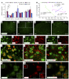
Figure 2. miR-186-5p is downregulated in the spinal cord after SNL and colocalized with CXCL13 in spinal neurons.
(A) The expression of miR-186-5p was decreased at days 1, 3, and 10 after SNL, while the expression of miR-1264-3p or miR-325-3p was not significantly changed after SNL. *P < 0.05; **P < 0.01, 1-way ANOVA followed by Bonferroni’s test. n = 3–6 mice/group. (B) Correlation analysis of the expression of Cxcl13 mRNA with the expression of miR-186-5p, miR-1264-3p, or miR-325-3p, respectively. The natural logarithm (LN) Cxcl13 mRNA expression in the spinal cord was negatively correlated with the expression of miR-186-5p. R2 = 0.8517; P < 0.05. Diamonds, squares, and triangles represent groups of individuals (mean value) across time. (C and D) FISH shows the expression of miR-186-5p in naive (C) and SNL (D) mice. Scale bar: 200 μm. (E and F) High magnification image of D, indicated in the white boxes of D. Scale bars: 100 μm. miR-186-5p was decreased in the ipsilateral dorsal horn (E, ipsi) compared with the contralateral dorsal horn (F, contra) 10 days after SNL. (GO) FISH for miR-186-5p combined with immunofluorescence staining for cell markers NeuN (GI), GFAP (JL), and Iba-1 (MO), respectively. Scale bars: 20 μm. miRNA-186-5p expression was only detected in NeuN-labeled neurons (I) in spinal dorsal horn. (PR) Combined FISH and immunofluorescence show that miR-186-5p (P) was colocalized with CXCL13 (Q and R). Scale bars: 10 μm; 5 μm (insets).
Figure 1
Figure 1. CXCL13 is upregulated in spinal neurons after SNL and contributes to SNL-induced neuropathic pain.
(A) The time course of Cxcl13 mRNA expression in the ipsilateral dorsal horn from naive (n = 6), sham, and SNL-operated mice. SNL increased Cxcl13 expression at 1 day (n = 3), 3 days (n = 5), 10 days (n = 3), and 21 days (n = 5) compared with sham 1 day (n = 5), 3 days (n = 3), 10 days (n = 3), and 21 days (n = 5), respectively. *P < 0.05; ***P < 0.001, Student’s t test. (B) ELISA shows increased CXCL13 protein levels in the ipsilateral dorsal horn after SNL (n = 4) compared with sham (n = 5) or naive (n = 5) animals. ***P < 0.001, Student’s t test. (C) ELISA shows CXCL13 protein increase in the CSF of mice with neuropathic pain (n = 9) compared with sham (n = 7) or naive mice (n = 8). *P < 0.05, Student’s t test. (DF) Representative images of CXCL13 immunofluorescence in the L5 dorsal horn. CXCL13 IR was low in naive mice (D), but increased in the ipsilateral dorsal horn (E) compared with the contralateral dorsal horn (F) 10 days after SNL. Scale bars: 100 μm. (GK) In situ hybridization of Cxcl13 mRNA and immunofluorescence staining of NeuN (GI), GFAP (J), and Iba-1 (K) shows that Cxcl13 mRNA was colocalized with neuronal marker, but not with markers of astrocytes or microglia. Scale bars: 50 μm; 10 μm (insets). (L) Double staining of CXCL13 and neuronal marker MAP2 in cultured dorsal horn neuron. Note the distribution of CXCL13 in neurite terminals (arrows). Scale bar: 25 μm. (M and N) Inhibition of Cxcl13 by shRNA lentivirus (n = 9) attenuated SNL-induced mechanical allodynia (M) and thermal hyperalgesia (N) compared with LV-NC treatment (n = 8). Intraspinal injection was performed 3 days after SNL (arrows). *P < 0.05; **P < 0.01; ***P < 0.001, 2-way repeated measures ANOVA followed by Bonferroni’s test.

References

    1. Charo IF, Ransohoff RM. The many roles of chemokines and chemokine receptors in inflammation. N Engl J Med. 2006;354(6):610–621. doi: 10.1056/NEJMra052723. - DOI - PubMed
    1. Asensio VC, Campbell IL. Chemokines in the CNS: plurifunctional mediators in diverse states. Trends Neurosci. 1999;22(11):504–512. doi: 10.1016/S0166-2236(99)01453-8. - DOI - PubMed
    1. Savarin-Vuaillat C, Ransohoff RM. Chemokines and chemokine receptors in neurological disease: raise, retain, or reduce? Neurotherapeutics. 2007;4(4):590–601. doi: 10.1016/j.nurt.2007.07.004. - DOI - PMC - PubMed
    1. Kiguchi N, Kobayashi Y, Kishioka S. Chemokines and cytokines in neuroinflammation leading to neuropathic pain. Curr Opin Pharmacol. 2012;12(1):55–61. doi: 10.1016/j.coph.2011.10.007. - DOI - PubMed
    1. Old EA, Malcangio M. Chemokine mediated neuron-glia communication and aberrant signalling in neuropathic pain states. Curr Opin Pharmacol. 2012;12(1):67–73. doi: 10.1016/j.coph.2011.10.015. - DOI - PubMed

Publication types