Clinical Trial

Molecular etiology of arthrogryposis in multiple families of mostly Turkish origin

Yavuz Bayram et al. J Clin Invest. 2016 Feb.

Abstract

Background: Arthrogryposis, defined as congenital joint contractures in 2 or more body areas, is a clinical sign rather than a specific disease diagnosis. To date, more than 400 different disorders have been described that present with arthrogryposis, and variants of more than 220 genes have been associated with these disorders; however, the underlying molecular etiology remains unknown in the considerable majority of these cases.

Methods: We performed whole exome sequencing (WES) of 52 patients with clinical presentation of arthrogryposis from 48 different families.

Results: Affected individuals from 17 families (35.4%) had variants in known arthrogryposis-associated genes, including homozygous variants of cholinergic γ nicotinic receptor (CHRNG, 6 subjects) and endothelin converting enzyme-like 1 (ECEL1, 4 subjects). Deleterious variants in candidate arthrogryposis-causing genes (fibrillin 3 [FBN3], myosin IXA [MYO9A], and pleckstrin and Sec7 domain containing 3 [PSD3]) were identified in 3 families (6.2%). Moreover, in 8 families with a homozygous mutation in an arthrogryposis-associated gene, we identified a second locus with either a homozygous or compound heterozygous variant in a candidate gene (myosin binding protein C, fast type [MYBPC2] and vacuolar protein sorting 8 [VPS8], 2 families, 4.2%) or in another disease-associated genes (6 families, 12.5%), indicating a potential mutational burden contributing to disease expression.

Conclusion: In 58.3% of families, the arthrogryposis manifestation could be explained by a molecular diagnosis; however, the molecular etiology in subjects from 20 families remained unsolved by WES. Only 5 of these 20 unrelated subjects had a clinical presentation consistent with amyoplasia; a phenotype not thought to be of genetic origin. Our results indicate that increased use of genome-wide technologies will provide opportunities to better understand genetic models for diseases and molecular mechanisms of genetically heterogeneous disorders, such as arthrogryposis.

Funding: This work was supported in part by US National Human Genome Research Institute (NHGRI)/National Heart, Lung, and Blood Institute (NHLBI) grant U54HG006542 to the Baylor-Hopkins Center for Mendelian Genomics, and US National Institute of Neurological Disorders and Stroke (NINDS) grant R01NS058529 to J.R. Lupski.


Figures

Figure 8
Figure 8. PSD3 variant showing an autosomal dominant inheritance identified in the patients with APS.
(A) Sanger sequencing analysis showing the segregation result of PSD3 variant identified in family HOU2061. (B) Elbow photographs of the affected individuals.
Figure 7
Figure 7. MYO9A variants identified in BAB6499.
(A and B) Segregation analyses of the identified MYO9A variants and patient photographs. While c.608A>G variant is inherited from the mother, c.6845G>A variant is observed as de novo. (C) Peptide alignment of human MYO9A with MYO9A in other species. Gly2282 and Tyr203 are conserved residues across all vertebrates. (D) Interactome of MYO9A (red circle) that shows the interactions with known arthrogryposis gene products (black circles). Gray circles indicate the proteins in the same network but not associated with arthrogryposis previously.
Figure 6
Figure 6. Variants in FBN3 identified in BAB7826.
(A) Family pedigree and segregation results of the identified compound heterozygous variants in FBN3. (B) Patient photographs showing the representative clinical sign of arthrogryposis.
Figure 5
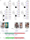
Figure 5. Homozygous VPS8 and POLR3A variants identified in BAB7143.
(A) Segregation analyses of the WES-detected variants. The proband was homozygous, and the parents were heterozygous carrier, consistent with Mendelian expectations. (B) Patient photographs showing clinical features, including ptosis, joint contractures, ulnar deviation of the hands, and surgically repaired foot deformity. (C) The interaction network of VPS8 (red circle) with other gene products, such as VPS33B, VPS53, and VIPAS39 (black circles), which were previously associated with different arthrogryposis-related disorders. Gray circles indicate the proteins in the same network but not associated with arthrogryposis previously. (D) Peptide alignment showing the conservation of the affected amino acids across the species.
Figure 4
Figure 4. Variants in GPR126 and MYBPC2 identified in patient BAB6212.
(A) Segregation analyses of the homozygous GPR126 and compound heterozygous MYBPC2 mutations in the family HOU2332. (B) Conservation alignment indicating that the affected amino acids of MYBPC2 are conserved across different vertebrates. (C) The interaction network of MYBPC2 (red circle) with known arthrogryposis gene products (black circles). Gray circles indicate the proteins in the same network but not associated with arthrogryposis previously.
Figure 3
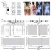
Figure 3. MYO18B and MYH7B variants identified in patient BAB7140.
(A) The pedigree of the HOU2620 family and segregation of the MYO18B and MYH7B variants. DNA sample of the mother was not available. (B) Patient photographs and X-ray images showing the severe phenotype most probably due to the synergistic effect of 2 homozygous mutations in 2 different arthrogryposis-related genes. Note the progressive scoliosis observed in the X-rays taken in 2012 and 2014, respectively. (C) Top panel: B-allele frequency plot of the entire chromosome 22 and 20, respectively. Bottom panel: Zoomed-in views of the regions encompassing MYO18B and MYH7B indicated by blue. AOH regions are shown as gray regions. Note the large AOH block that nearly encompass entire chromosome 20, suggesting a possible uniparental disomy.
Figure 2
Figure 2. Segregation results, photographs, and variant distributions of the patients with CHRNG mutations.
(A) Pedigrees and Sanger sequencing analyses of the patients that represent the proper segregation of the WES-detected CHRNG variants. One of the patients with more severe phenotype (BAB5611) has another homozygous variant in a known arthrogryposis gene (ERCC2), which also segregates in the family. (B) Photographs of the probands showing the major clinical features, including joint contractures, multiple pterygiums, and distinct facial dysmorphism. (C and D) Schematic representations of gene and protein structures of CHRNG and localization of the identified variants. Neur_chan_LBD, neurotransmitter-gated ion-channel ligand binding domain; Neur_chan_memb, neurotransmitter-gated ion-channel transmembrane region.
Figure 1
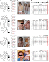
Figure 1. Segregation analyses, photographs, and AOH regions of the patients with ECEL1 mutations.
(A) Detailed pedigrees of the families and their corresponding Sanger-sequencing chromatograms showing the segregation analyses. (B) Photographs of the patients. Note the whistling-face appearance of BAB6500, which is commonly observed in DA type 2A rather than ECEL1-associated DA type 5D. (C) AOH study of the patients based on calculated B-allele frequency data culled from WES analysis. Gray shaded areas indicate AOH regions. Note the approximately 840-Kb overlap between the AOH regions, which includes the ECEL1 gene.

References

    1. Lowry RB, Sibbald B, Bedard T, Hall JG. Prevalence of multiple congenital contractures including arthrogryposis multiplex congenita in Alberta, Canada, and a strategy for classification and coding. Birth Defects Res A Clin Mol Teratol. 2010;88(12):1057–1061. doi: 10.1002/bdra.20738. - DOI - PubMed
    1. Hall JG. Arthrogryposis (multiple congenital contractures): diagnostic approach to etiology, classification, genetics, and general principles. Eur J Med Genet. 2014;57(8):464–472. doi: 10.1016/j.ejmg.2014.03.008. - DOI - PubMed
    1. Filges I, Hall JG. Failure to identify antenatal multiple congenital contractures and fetal akinesia — proposal of guidelines to improve diagnosis. Prenat Diagn. 2013;33(1):61–74. doi: 10.1002/pd.4011. - DOI - PubMed
    1. Haliloglu G, Topaloglu H. Arthrogryposis and fetal hypomobility syndrome. Handb Clin Neurol. 2013;113:1311–1319. doi: 10.1016/B978-0-444-59565-2.00003-4. - DOI - PubMed
    1. Bamshad M, Jorde LB, Carey JC. A revised and extended classification of the distal arthrogryposes. Am J Med Genet. 1996;65(4):277–281. doi: 10.1002/(SICI)1096-8628(19961111)65:4<277::AID-AJMG6>3.0.CO;2-M. - DOI - PubMed

Publication types