

Nonenhancing Leptomeningeal Metastases: Imaging Characteristics and Potential Causative Factors
- PMID: 26753054
- PMCID: PMC4680901
- DOI: 10.1177/1941874415591702
Nonenhancing Leptomeningeal Metastases: Imaging Characteristics and Potential Causative Factors
Abstract
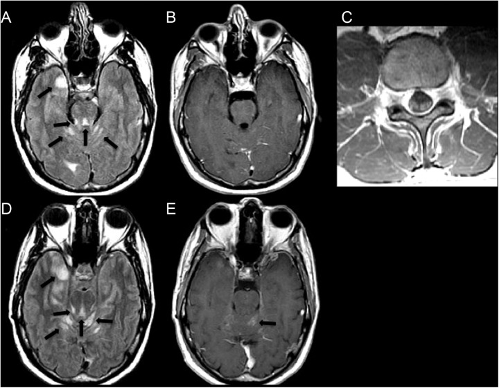
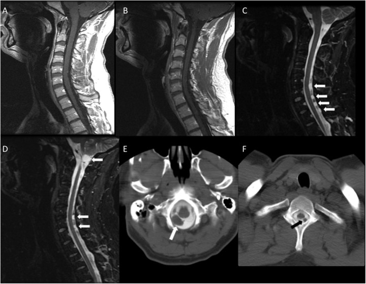
The diagnosis of leptomeningeal metastasis (LM) has increased in frequency, as new therapies have lengthened the survival of patients with cancer. Early diagnosis and intervention help improve quality of life and prevent further neurological deterioration in LM. The detection of LM is often established by magnetic resonance imaging examinations, cerebrospinal fluid analysis, or both. We present a series of cases where LM was identified on fluid-attenuated inversion recovery or T2-weighted image but was nonenhancing on the traditionally more sensitive postcontrast T1-weighted sequences. Nonenhancing LM is unusual and not yet fully understood but should be considered in the appropriate clinical context and may become more common with increased utilization of antiangiogenic therapies.
Keywords: brain neoplasms; imaging; meningeal neoplasms; nervous system neoplasms; techniques.
Conflict of interest statement
Figures
Similar articles
-
Relationship between contrast enhancement on fluid-attenuated inversion recovery MR sequences and signal intensity on T2-weighted MR images: visual evaluation of brain tumors.J Magn Reson Imaging. 2005 Jun;21(6):694-700. doi: 10.1002/jmri.20331. J Magn Reson Imaging. 2005. PMID: 15906343
-
Leptomeningeal metastases.Neurol Clin. 2003 Feb;21(1):25-66. doi: 10.1016/s0733-8619(02)00032-4. Neurol Clin. 2003. PMID: 12690644 Review.
-
Classification of Leptomeningeal Metastases from Solid Organ Malignancies and Clinical Outcomes: Series from a Cancer Research Centre.Indian J Surg Oncol. 2020 Jun;11(2):308-312. doi: 10.1007/s13193-020-01071-0. Epub 2020 Apr 17. Indian J Surg Oncol. 2020. PMID: 32523280 Free PMC article.
-
Contrast-enhanced 3-dimensional Fluid-attenuated Inversion Recovery Sequences Have Greater Sensitivity for Detection of Leptomeningeal Metastases in Pediatric Brain Tumors Compared With Conventional Spoiled Gradient Echo Sequences.J Pediatr Hematol Oncol. 2018 May;40(4):316-319. doi: 10.1097/MPH.0000000000000957. J Pediatr Hematol Oncol. 2018. PMID: 28902082 Clinical Trial.
-
New approaches to and current treatment of leptomeningeal metastases.Curr Opin Neurol. 1994 Dec;7(6):492-500. doi: 10.1097/00019052-199412000-00004. Curr Opin Neurol. 1994. PMID: 7532525 Review.
Cited by
-
Leptomeningeal Metastasis: The Role of Cerebrospinal Fluid Diagnostics.Front Neurol. 2019 Aug 20;10:839. doi: 10.3389/fneur.2019.00839. eCollection 2019. Front Neurol. 2019. PMID: 31481919 Free PMC article.
-
Extraordinary linear non-enhancing brainstem leptomeningeal metastasis from lung adenocarcinoma: A case report.Clin Case Rep. 2024 May 14;12(5):e8932. doi: 10.1002/ccr3.8932. eCollection 2024 May. Clin Case Rep. 2024. PMID: 38751962 Free PMC article.
-
Intra-operative cerebrospinal fluid sampling versus post-operative lumbar puncture for detection of leptomeningeal disease in malignant paediatric brain tumours.PLoS One. 2018 May 3;13(5):e0196696. doi: 10.1371/journal.pone.0196696. eCollection 2018. PLoS One. 2018. PMID: 29723241 Free PMC article.
-
T2-hyperintense, non-gadolinium-enhancing, diffusion-restricted symmetrical curvilinear lesion of the anterior pons: a rare radiological pattern of leptomeningeal metastatic disease secondary to likely lung adenocarcinoma.BMJ Case Rep. 2021 Jan 18;14(1):e236265. doi: 10.1136/bcr-2020-236265. BMJ Case Rep. 2021. PMID: 33461996 Free PMC article.
-
Band-Like Brainstem Lesion in a Patient With a History of Lung Adenocarcinoma.Cureus. 2022 Oct 26;14(10):e30726. doi: 10.7759/cureus.30726. eCollection 2022 Oct. Cureus. 2022. PMID: 36320789 Free PMC article.
References
-
- Groves MD. Leptomeningeal disease. Neurosurg Clin N Am. 2011;22:67–78. - PubMed
-
- Singh SK, Agris JM, Leeds NE, Ginsberg LE. Intracranial leptomeningeal metastases: Comparison of depiction at FLAIR and contrast-enhanced MR imaging. Radiology. 2000;217:50–3. - PubMed
-
- Stuckey SL, Goh TD, Heffernan T, Rowan D. Hyperintensity in the subarachnoid space on FLAIR MRI. AJR Am J Roentgenol. 2007;189:913–21. - PubMed
LinkOut - more resources
Full Text Sources
Other Literature Sources