Review
. 2015 Dec 30:4:835.
doi: 10.1186/s40064-015-1604-4. eCollection 2015.

Does malalignment affect revision rate in total knee replacements: a systematic review of the literature

Affiliations
Review

Does malalignment affect revision rate in total knee replacements: a systematic review of the literature

Mohammed Hadi et al. Springerplus. .

Abstract

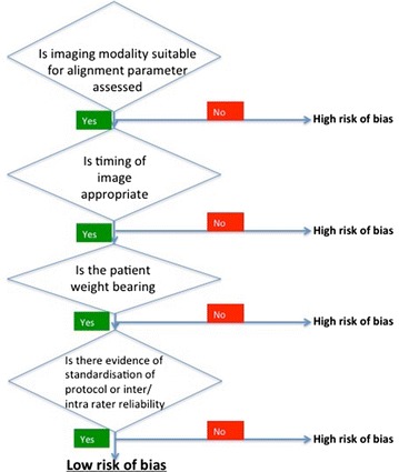
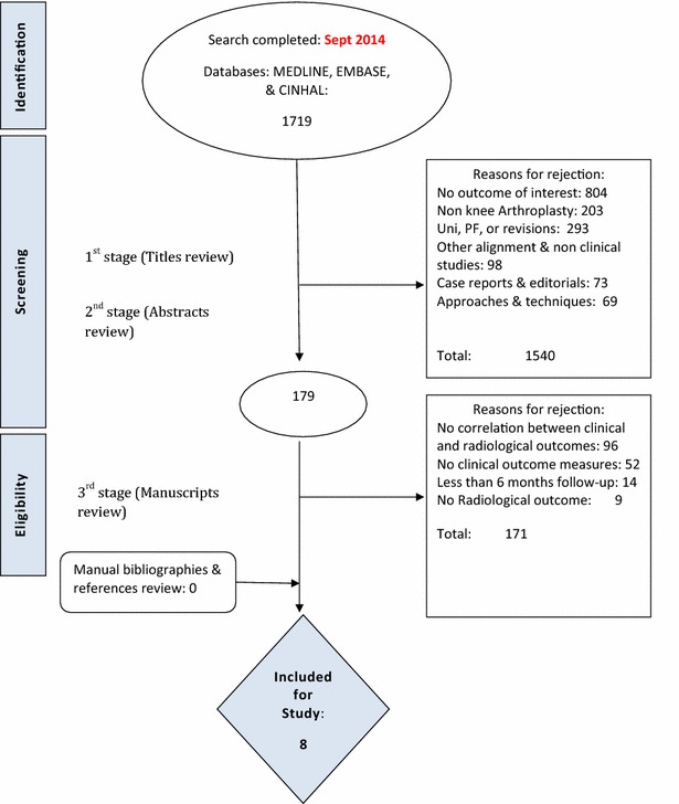
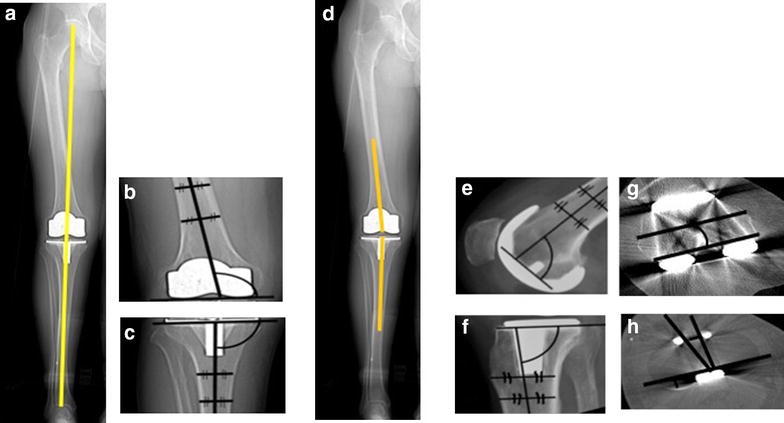
To ensure implant durability following Modern total knee replacement (TKR) surgery, one long held principle in condylar total knee arthroplasty is positioning the components in alignment with the mechanical axis and restoring the overall limb alignment to 180° ± 3°. However, this view has been challenged recently. Given the high number of TKR performed, clarity on this integral aspect of the procedure is necessary. To investigate the association between malalignment following primary TKR and revision rates. A systematic review of the literature was conducted using a computerised literature search of Medline, CINHAL, and EMBASE to identify English-language studies published from 2000 through to 2014. Studies with adequate information on the correlation between malalignment and revision rate with a minimum follow-up of 6 months were considered for inclusion. A study protocol, including the detailed search strategy was published on the PROSPERO database for systematic reviews. From an initial 2107 citations, eight studies, with variable methodological qualities, were eligible for inclusion. Collectively, nine parameters of alignment were studied, and 20 assessments were made between an alignment parameter and revision rate. Four out of eight studies demonstrated an association between a malalignment parameter and increased revision rates. In the coronal plane, only three studies assessed the mechanical axis. None of these studies found an association with revision rates, whereas four of the five studies investigating the anatomical axis found an association between malalignment and increased revision rate. This study demonstrates the effect of malalignment on revision rates is likely to be modest. Interestingly, studies that used mechanical alignment in the coronal plane demonstrated no association with revision rates. This questions the premise of patient specific instrumentation devices based on the mechanically aligned knee when considering revision as the endpoint.

Keywords: Malalignment; Revision rate; Systematic review; Total knee arthroplasty.


Figures

Fig. 1
Fig. 1
Radiological assessment quality (RAQ) criteria for assessing alignment. The evaluation was done using a five yes/no question checklist that was devised for this review. A sensitivity analysis was performed to determine if the quality of the radiological methods was an important factor in the outcome. The rationale for each set of questions was as follows: The suitability of the imaging modality used: Overall limb alignment is better assessed on a whole leg radiograph compared to a short film radiographs (Moreland 1988) and Short film x-rays are used for the assessment of component’s anatomical alignment (Morgan et al. 2008). The timing of the imaging: Malalignment on images acquired several years following surgery may be secondary to implant subsidence/migration (Morgan et al. 2008). The patient’s weight bearing status at the time of imaging: the relationship between the bony and soft tissue parts of the knee joint is most visible during stressing manoeuvre such as weight bearing (Nicoll and Rowley 2010). Indication of standardisation when acquiring the images: Non-standardised protocols for acquiring images can result in inconsistent magnification and rotation, introducing a source of bias (Parratte et al. ; Registry 2013). Evidence of rater reliability when assessing the images for alignment: To ensure consistency (Parratte et al. ; Registry 2013)
Fig. 2
Fig. 2
PRISMA flow diagram including the details of our search results for this review. Figure shows the reasons behind study exclusion at each stage of the search and the number of studies identified at each point of the search
Fig. 3
Fig. 3
A diagrammatic representation of different alignment parameters based on the knee society total knee arthroplasty roentgenographic evaluation and scoring system24. The coronal tibiofemoral mechanical angle is the angle resulting from drawing a line from the centre of the femoral head down to centre of the ankle through the centre of the knee (a)—ideally 180°. The coronal femoral angle cFA (b)—ideally 96°—and coronal tibial angle cTA (c)—ideally 90°—are the angles between the components’ coronal axes (the line connecting the femoral components most distal condyles and the line along the horizontal tibial plate) and the bones’ coronal anatomical axes (line which bisects the medullary canal of the femur and tibia respectively). The coronal tibiofemoral anatomical angle is a combination of the coronal anatomical femoral axis and coronal anatomical tibial axis (d). The sagittal femoral sFA (e)—ideally 90°—and sagittal tibial sTA (f)— ideally between 83 and 90°—angles are the angles between the components’ sagittal axes (horizontal line perpendicular to the femoral component peg and line along the horizontal tibial plate) and the anatomical sagittal bones’ axes (line which bisects the medullary canal of the femur and tibia respectively). The axial femoral (aFRA) (g)—ideally 0°—and axial tibial—ideally within 15°—(aTRA) (h) angles are the angles between the components’ axial axes (line through the centre of the femoral pegs and the line through the most posterior points of the tibial plate on axial views respectively) and the bones’ axial axes (surgical epicondylar femoral axis and the tibial tuberosity axis respectively). The combined components axial (aCRA) rotational alignment angles—ideally 0°—is the angle between the components axial axes

References

    1. Bach CM, Mayr E, Liebensteiner M, et al. Correlation between radiographic assessment and quality of life after total knee arthroplasty. Knee. 2009;16:207–210. doi: 10.1016/j.knee.2008.11.003. - DOI - PubMed
    1. Bargren JH, Blaha JD, Freeman MA. Alignment in total knee arthroplasty. Correlated biomechanical and clinical observations. Clin Orthop Relat Res. 1983;173:178–183. - PubMed
    1. Berend ME, Ritter MA, Hyldahl HC, et al. Implant migration and failure in total knee arthroplasty is related to body mass index and tibial component size. J Arthroplasty. 2008;23:104–109. doi: 10.1016/j.arth.2008.05.020. - DOI - PubMed
    1. Berend ME, Ritter MA, Meding JB, et al. Tibial component failure mechanisms in total knee arthroplasty. Clin Orthop Relat Res. 2004;428:26–34. doi: 10.1097/01.blo.0000148578.22729.0e. - DOI - PubMed
    1. Bhandari M, Chiavaras MM, Parasu N, et al. Radiographic union score for hip substantially improves agreement between surgeons and radiologists. BMC Musculoskelet Disord. 2013;14:70. doi: 10.1186/1471-2474-14-70. - DOI - PMC - PubMed

LinkOut - more resources