

Case Reports
Epub 2015 Dec 22.
Ultrasound and multidetector computed tomography of mandibular salivary gland adenocarcinoma in two dogs
Affiliations
- PMID: 26753133
- PMCID: PMC4703874
Item in Clipboard
Case Reports
Ultrasound and multidetector computed tomography of mandibular salivary gland adenocarcinoma in two dogs
Open Vet J.
2015.
Abstract
Malignant tumors of the salivary glands are rare in dogs, with adenocarcinoma being the most represented. Parotid and mandibular glands are most commonly affected in dogs. Because of local invasivity and high metastatic potential, preoperative imaging evaluation of mandibular region and tumoral staging is essential along with biopsy sampling. The present manuscript describes the ultrasound and computed tomographic imaging findings of mandibular gland adenocarcinoma in two dogs and discusses their clinical utility.
Keywords: Computed tomography; Dog; Salivary gland tumor; Ultrasound.
Conflict of interest statement
Figures
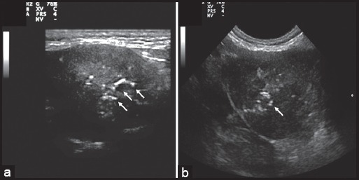
Dog 1. Longitudinal (a) and transversal (b) ultrasound scans of the left submandibular gland - ventral approach. There is enlargement of the gland with a hinomogeneous, hypo echoic parenchyma and small hyperechoic calcific foci (arrows).

Dog 1. Unenhanced (a) and contrast-enhanced (b) transverse comptuted tomography images of the neoplastic left mandibular gland. Oblique Dorsal multiplanar reformatted CT images at level of mandibular glands (c) and medial retropharyngeal lymphnodes (d). The affected gland appeared enlarged compared to the contralateral one (c) with parenchimal mineralizations (black arrows) and small hypoattenuating areas suggestive of necrosis (b, arrowhead). The left medial retropharyngeal lymph node (b, single arrow) was enlarged compared with its contralateral (d, triple arrows). Rmg: right mandibular gland; lmg: left mandibular gland

Dog 2. Transverse ultrasound scans of the right submandibular gland, before (a) and after (b) ultrasound guided drainage - ventral approach. The gland is enlarged with anhecoic fluid filled cavity (a, asterisk). The solid component shows heterogeneous echogenicity with small hyperechoic calcific foci (arrows). Residual intraparenchimal fluid (asterisk) and calcific foci (arrow) are seen after drainage of the fluid component (b).

Dog 2. Volume rendered computed tomography images of the left (a) and right (b) mandibular region. Unenhanced (c) and contrast-enhanced (d) transverse comptuted tomography images of the neoplastic right mandibular gland. The affected gland (rm) appeared as a cavitated mass with omogeneous hypoattenuating fluid content and focal mineralization (arrows). The enhancement of peripheral parenchymal component was poor. Volume rendered image of the right mandibular region (b) clarified the relationships between the neoplastic gland and surrounding structures especially if compared with the normal anatomy of the left side (a). The mass displaced dorsally the parotid gland (p) and compressed the retromandibular vein (rv), facial vein (fv) and mandibular lymph nodes (lf). No sign of muscular or bone infiltration were evident. g: jugular vein; lm: left mandibular gland; dm: digastric muscle.

Dog 2. (a) Oblique dorsal multiplanar reformatted contrast-enhanced CT image at level of medial retropharyngeal lymph nodes. Transverse contrast-enhanced CT image at the level of cranial lobe (b), middle part (c) and caudal lobe of the left lung. Right medial retropharyngeal lymph node (rrl) was enlarged compared to its contralateral (lrl). In different parts of the left lung multiple round nodular lesions isoattenuating to soft tissue, were present, compatible with pulmonary metastasis. mg: right mandibular gland – dorsal margin; rlm: right longus capitis muscle; llm: left longus capitis muscle; e: esophagus.
References
-
- Almeida A.P, Malm C, Lavalle G, Cassali G.D, Santos R.L, Paixão T.A. Salivary gland carcinosarcoma in a dog. Braz. J. Vet. Pathol. 2010;3:137–141.
-
- Burek D.A, Munn R.J, Madewell B.R. Metastatic adenocarcinoma of a minor0020salivary gland in a cat. Zentralbl. Veterinarmed. A. 1994;41:485–490. - PubMed
-
- Burns G.O, Scrivani P.V, Thompson M.S, Erb H.N. Relation between age, body weight, and medial retropharyngeal lymph node size in apparently healthy dogs. Vet. Radiol. Ultrasound. 2008;49:277–281. - PubMed
-
- Cannon M.S, Paglia D, Zwingenberger A.L, Boroffka S.A, Hollingsworth S.R, Wisner E.R. Clinical and diagnostic imaging findings in dogs with zygomatic sialadenitis:11 cases (1990-2009) J. Am. Vet. Med. Assoc. 2011;239:1211–1218. - PubMed
-
- Carberry C.A, Flanders J.A, Harvey H.J, Ryan A.M. Salivary gland tumors in dogs and cats: a literature and case review. J. Am. Anim. Hosp. Assoc. 1988;24:561–567.
Publication types
LinkOut - more resources
Full Text Sources